. 2014 Aug 14:12:65.
doi: 10.1186/s12915-014-0065-5.

Biofilms formed by the archaeon Haloferax volcanii exhibit cellular differentiation and social motility, and facilitate horizontal gene transfer

Biofilms formed by the archaeon Haloferax volcanii exhibit cellular differentiation and social motility, and facilitate horizontal gene transfer

Scott Chimileski et al. BMC Biol. .

Abstract

Background: Archaea share a similar microbial lifestyle with bacteria, and not surprisingly then, also exist within matrix-enclosed communities known as biofilms. Advances in biofilm biology have been made over decades for model bacterial species, and include characterizations of social behaviors and cellular differentiation during biofilm development. Like bacteria, archaea impact ecological and biogeochemical systems. However, the biology of archaeal biofilms is only now being explored. Here, we investigated the development, composition and dynamics of biofilms formed by the haloarchaeon Haloferax volcanii DS2.

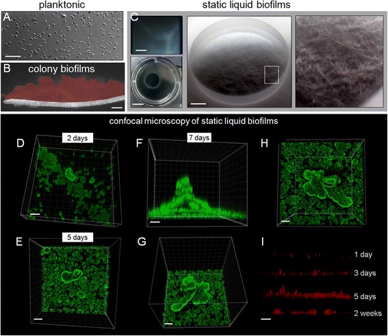
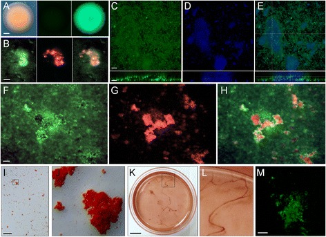
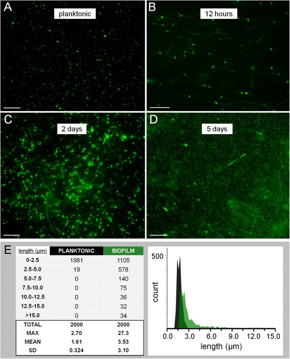
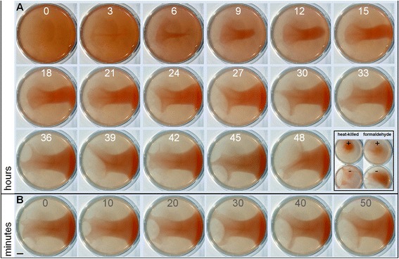
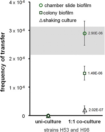
Results: Biofilms were cultured in static liquid and visualized with fluorescent cell membrane dyes and by engineering cells to express green fluorescent protein (GFP). Analysis by confocal scanning laser microscopy showed that H. volcanii cells formed microcolonies within 24 h, which developed into larger clusters by 48 h and matured into flake-like towers often greater than 100 μm in height after 7 days. To visualize the extracellular matrix, biofilms formed by GFP-expressing cells were stained with concanavalin A, DAPI, Congo red and thioflavin T. Stains colocalized with larger cellular structures and indicated that the extracellular matrix may contain a combination of polysaccharides, extracellular DNA and amyloid protein. Following a switch to biofilm growth conditions, a sub-population of cells differentiated into chains of long rods sometimes exceeding 25 μm in length, compared to their planktonic disk-shaped morphology. Time-lapse photography of static liquid biofilms also revealed wave-like social motility. Finally, we quantified gene exchange between biofilm cells, and found that it was equivalent to the mating frequency of a classic filter-based experimental method.

Conclusions: The developmental processes, functional properties and dynamics of H. volcanii biofilms provide insight on how haloarchaeal species might persist, interact and exchange DNA in natural communities. H. volcanii demonstrates some biofilm phenotypes similar to bacterial biofilms, but also has interesting phenotypes that may be unique to this organism or to this class of organisms, including changes in cellular morphology and an unusual form of social motility. Because H. volcanii has one of the most advanced genetic systems for any archaeon, the phenotypes reported here may promote the study of genetic and developmental processes in archaeal biofilms.


Figures

Figure 1
Figure 1
Growth and development of Haloferax volcanii static liquid biofilms. (A) Cells within typical shaken culture of H. volcanii DS2 under transmitted light. Scale bar equals 10 μm. (B) Cross section of cryo-processed H. volcanii colony biofilm grown on CA medium for 5 days and stained with CellMask Orange (CMO). Scale bar equals 30 μm. (C) Photographs of SL-biofilms routinely grown and analyzed within chamber slides (top left; scale bar equals 2 mm) and on glass coupons within six-well plates (bottom left; scale bar equals 1 cm). A macroscopic photograph of biofilm growth on a 12.7-mm glass coupon is shown in the center (scale bar equals 2 mm) with an area magnified on the right (shown in white box). (D-H) CLSM of biofilms grown on glass coupons (within a six-well plate in Hv-Ca medium; scale bars equal 30 μm). Biofilms stained with FM 1-43 were imaged directly in bulk Hv-starve medium using a 63× water-immersion objective after 2 days (D), 5 days (E) and 7 days (F-H). (I) Biofilm development shown through orthogonal views of SL-biofilms on glass coupons stained with CMO. CLSM, confocal laser scanning microscopy; CMO, CellMask Orange; SL-biofilm, static liquid biofilm.
Figure 2
Figure 2
Visualizing the extracellular matrix of Haloferax volcanii biofilms. (A) Development of a GFP-expressing strain for use in biofilm visualization: colonies formed by the parental strain H. volcanii H1206 under transmitted light (left), under blue excitation (center), and by the strain H. volcanii H1206(pJAM1020) under blue excitation (right). Scale bar equals 250 μm. (B) Seven-day H. volcanii H1206(pJAM1020) SL-biofilm stained with concanavalin A-Texas Red collected with blue excitation (left), green excitation (center), and shown as an overlay (right). Scale bar equals 20 μm. (C,D,E) Top-down projections of a Z-stack for a H. volcanii H1206(pJAM1020) SL-biofilm stained with DAPI. (C) GFP signal under blue excitation. (D) DAPI-stained material with violet excitation. (E) Overlay of (C) and (D). Orthogonal views from CLSM analysis are shown below each panel and the plane of the orthoslice is shown in (E). Scale bars equal 20 μm. (F,G,H) Seven-day H. volcanii H1206(pJAM1020) SL-biofilm stained with Congo red with blue excitation (F), and green excitation for CR fluorescence (G), with an overlay of (F) and (G) shown in (H). Scale bar equals 20 μm. (I) Bright-field view of 7-day CR-stained SL-biofilm within a chamber slide. Scale bar equals 1 mm. Area outlined by black box is shown in (J). (K,L) Ten-day SL-biofilm within Petri dish grown at 25°C in medium containing CR with CR-stained string or web-like structures magnified in (L). Scale bar equals 1 cm. (M) Seven-day H1206 SL-biofilms grown in Hv-YPC medium stained with thioflavin T under blue excitation (top down 3D projection of a Z-stack; scale bar equals 20 μm). CLSM, confocal laser scanning microscopy; CR, Congo red; DAPI, 4′,6-diamidino-2-phenylindole; GFP, green fluorescent protein; SL-biofilm, static liquid biofilm.
Figure 3
Figure 3
Cellular morphology within developing Haloferax volcanii H1206(pJAM1020) static liquid biofilms. (A) Planktonic H. volcanii H1206(pJAM1020) cells from exponential phase shaking Hv-YPC culture. (B,C,D) Cells within a developing H1206(pJAM1020) SL-biofilm grown in a chamber slide (in Hv-YPC medium) after 12 h (B), 2 days (C) and 5 days (D). (E) Table listing the number of cells binning into 2.5-μm categories and summarized statistics for 2,000 cells measured for planktonic and biofilm cell populations (left) and histogram showing distribution of length at 0.2-μm binning intervals for planktonic (black) and biofilm (green) cells (right). Cell length was measured with Fiji particle analysis using images collected from three independent exponential phase shaking cultures and three 12 h biofilms grown in chamber slides. Mean length of populations was statistically different with P < 0.0001. Scale bars equal 20 μm. SD, standard deviation; SL-biofilm, static liquid biofilm.
Figure 4
Figure 4
Time-lapse macroscopic photography of static liquid biofilm reformation. An established 7-day SL-biofilm grown in a plastic Petri dish in rich medium (Hv-YPC) was mechanically homogenized and left to incubate at 42°C while photographs were taken at regular time intervals. (A) Biofilm reformation over a 2-day period, with images shown at 3 h intervals. Inlay: Cellular vitality controls shown as still images from time-lapse series (see Additional files 6 and 7: Movies 5 and 6) of a SL-biofilm treated with heat (left) and with 4% formaldehyde (right). (B) SL-biofilm imaged at 10-min intervals over a 50-min period (a series from between 45 h and 48 h above). Scale bar equals 1 cm. SL-biofilm, static liquid biofilm.
Figure 5
Figure 5
Recombination within Haloferax volcanii biofilms. Two double auxotrophic strains (H53, ∆pyrE2/∆trpA; H98, ∆pyrE2/hdrB) were mixed at equal cell densities and grown together in Hv-YPC + thymidine medium for 7 days under shaking conditions (black triangles), or as colony biofilms (green squares) and chamber slide SL-biofilms (green circles). Cultures and biofilms were then harvested, washed and plated on defined medium with uracil alone, to select for recombinants, which are either H53 cells that have regained tryptophan prototrophy through a transfer event with a H98 cell(s) or H98 cells that have gained thymidine prototrophy from a H53 cell(s). Average frequency of transfer is shown for three replicates per condition. The transfer frequency range reported in the study in which this HGT mechanism was discovered, traditionally conducted using nitrocellulose filters and known as mating, is shown as a grey box [56]. Vertical bars equal one standard deviation. HGT, horizontal gene transfer.

References

    1. Aguilar C, Vlamakis H, Losick R, Kolter R. Thinking about Bacillus subtilis as a multicellular organism. Curr Opin Microbiol. 2007;10:638–643. doi: 10.1016/j.mib.2007.09.006. - DOI - PMC - PubMed
    1. Watnick P, Kolter R. Biofilm, city of microbes. J Bacteriol. 2000;182:2675–2679. doi: 10.1128/JB.182.10.2675-2679.2000. - DOI - PMC - PubMed
    1. Parsek MR, Greenberg EP. Sociomicrobiology: the connections between quorum sensing and biofilms. Trends Microbiol. 2005;13:27–33. doi: 10.1016/j.tim.2004.11.007. - DOI - PubMed
    1. Branda SS, Vik S, Friedman L, Kolter R. Biofilms: the matrix revisited. Trends Microbiol. 2005;13:20–26. doi: 10.1016/j.tim.2004.11.006. - DOI - PubMed
    1. Flemming HC, Wingender J. The biofilm matrix. Nat Rev Microbiol. 2010;8:623–633. - PubMed

Publication types

LinkOut - more resources