Review
. 2014 Sep;297(9):1694-705.
doi: 10.1002/ar.22974.

Finding the sweet spot: assembly and glycosylation of the dystrophin-associated glycoprotein complex

Affiliations
Review

Finding the sweet spot: assembly and glycosylation of the dystrophin-associated glycoprotein complex

Dewayne Townsend. Anat Rec (Hoboken). 2014 Sep.

Abstract

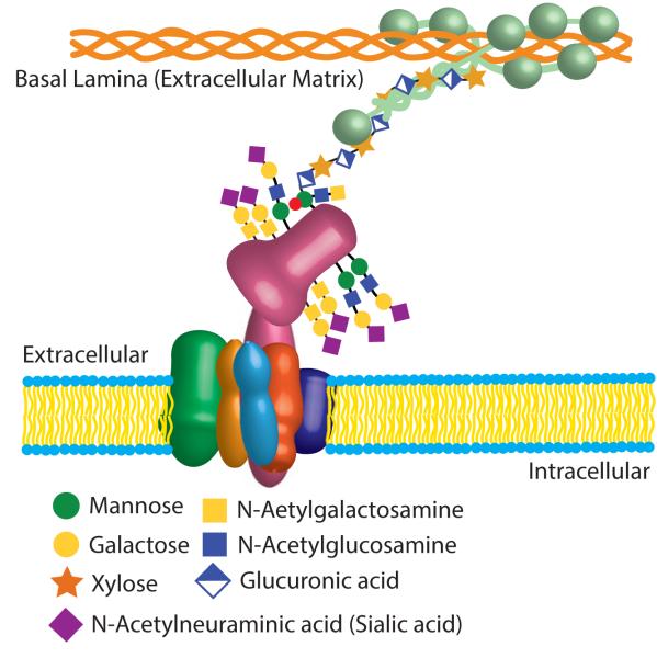
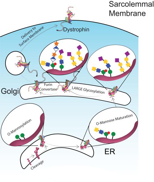
The dystrophin-associated glycoprotein complex (DGC) is a collection of glycoproteins that are essential for the normal function of striated muscle and many other tissues. Recent genetic studies have implicated the components of this complex in over a dozen forms of muscular dystrophy. Furthermore, disruption of the DGC has been implicated in many forms of acquired disease. This review aims to summarize the current state of knowledge regarding the processing and assembly of dystrophin-associated proteins with a focus primarily on the dystroglycan heterodimer and the sarcoglycan complex. These proteins form the transmembrane portion of the DGC and undergo a complex multi-step processing with proteolytic cleavage, differential assembly, and both N- and O-glycosylation. The enzymes responsible for this processing and a model describing the sequence and subcellular localization of these events are discussed.

Keywords: dystroglycan; dystrophin associated glycoprotein complexes; muscle; sarcoglycan; sarcospan.


Figures

Figure 1
Figure 1
A schematic cartoon of the DGC in the sarcolemmal membrane. The sarcoglycan complex (SGC) consists of α-SG (green), β-SG (orange), γ-SG (gold), σ-SG (light blue), and sarcospan (purple). This protein complex associates with β-dystroglycan (DG; pink) which binds to α-DG. α-DG is heavily glycosylated and functions as a receptor for laminin (light green), which in turn binds to collagen (orange). All of the components of the DGC shown here are also N-glycosylated; these features are not shown, but details can be found in the text.
Figure 2
Figure 2
A model of the processing of dystroglycan (DG; pink). The DAG polypeptide is cleaved into α- and β-DG. While shown in the ER, the nature of the protease and its precise subcellular location are unknown. O-linked mannosylation (green circles) of α-DG occurs within the ER. During O-mannose maturation, mannose moieties on which the LARGE-glycan will be added are phosphorylated (red circle) and N-acetylglucosamine (filled blue square) and N-acetylgalactosamine (filled yellow square) are added. Following transit to the Golgi, additional O-glycosylation occurs with many structures capped with galactose (yellow circle) and sialic acid (purple diamond). Next a polymer of glucuronic acid (half-filled diamond) and xylose (orange star) is added by the glycosyltransferase LARGE. Following the cleavage of the N-terminal globular domain of α-DG by a furin convertase the complex is exported to the sarcolemmal membrane. The number of sites and structures of the glycosylation moieties has been simplified for clarity (see text for more details). Note the subcellular location where dystrophin first interacts with the DGC is not clear, while evident at the membrane so data suggests that dystrophin has a role in exporting the DGC from the Golgi (see text for details).

References

    1. Acharyya S, Butchbach MER, Sahenk Z, Wang H, Saji M, Carathers M, Ringel MD, Skipworth RJE, Fearon KCH, Hollingsworth MA, Muscarella P, Burghes AHM, Rafael-Fortney JA, Guttridge DC. Dystrophin glycoprotein complex dysfunction: a regulatory link between muscular dystrophy and cancer cachexia. Cancer Cell. 2005;8:421–432. - PubMed
    1. Adams ME, Mueller HA, Froehner SC. In vivo requirement of the alpha-syntrophin PDZ domain for the sarcolemmal localization of nNOS and aquaporin-4. J Cell Biol. 2001;155:113–122. - PMC - PubMed
    1. Ahn AH, Yoshida M, Anderson MS, Feener CA, Selig S, Hagiwara Y, Ozawa E, Kunkel LM. Cloning of human basic A1, a distinct 59-kDa dystrophin-associated protein encoded on chromosome 8q23-24. Proc Natl Acad Sci U S A. 1994;91:4446–4450. - PMC - PubMed
    1. Albrecht DE, Froehner SC. DAMAGE, a novel alpha-dystrobrevin-associated MAGE protein in dystrophin complexes. J Biol Chem. 2004;279:7014–7023. - PubMed
    1. Alhamidi M, Kjeldsen Buvang E, Fagerheim T, Brox V, Lindal S, Van Ghelue M, Nilssen O. Fukutin-related protein resides in the Golgi cisternae of skeletal muscle fibres and forms disulfide-linked homodimers via an N-terminal interaction. PLoS One. 2011;6:e22968. - PMC - PubMed

Publication types

MeSH terms

LinkOut - more resources