Randomized Controlled Trial
. 2014:125:27-42; discussion 42-4.

Translational studies in older men using testosterone to treat sarcopenia

Affiliations
Randomized Controlled Trial

Translational studies in older men using testosterone to treat sarcopenia

Randall J Urban et al. Trans Am Clin Climatol Assoc. 2014.

Abstract

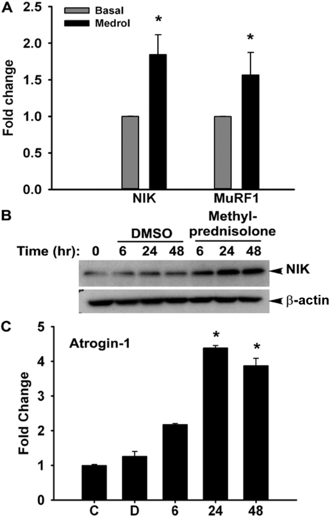
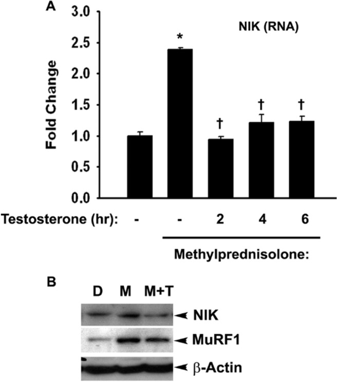
Sarcopenia is the loss of skeletal muscle mass and strength that occurs with aging. Our research group has found an efficacious administration paradigm using testosterone to combat sarcopenia in humans. In addition, our research has uncovered an important regulatory enzyme of inflammation, nuclear factor-κB-inducing kinase that may regulate human skeletal muscle catabolism, and that appears to be counter-regulated by administration of standard doses of testosterone. This is important because a number of age-related clinical circumstances trigger acute and chronic muscle loss including cancer, chronic obstructive pulmonary disease, hospitalization, acute and chronic illness, and diseases in which systemic inflammation occurs. Moreover, it is often the treatment itself that can induce muscle loss. For example, glucocorticoids are tremendously effective at reducing inflammation and are a frontline therapy for many inflammatory-based diseases, yet paradoxically trigger muscle loss. We will discuss our research findings and the clinical significance of our human clinical translational research with testosterone.


Conflict of interest statement

Potential Conflicts of Interest: None Disclosed.

Figures

Fig. 1
Fig. 1
Increased muscle NIK mRNA levels in old subjects. Human vastus lateralis muscle was sampled from young (29 ± 6 years) vs older (67 ± 5 years) subjects and assayed for NIK expression using qRT-PCR, with data analyzed using the delta CT method with GAPDH as the reference standard (lower delta CT means higher mRNA expression).
Fig. 2
Fig. 2
Decrease in skeletal muscle NIK message and protein levels after testosterone therapy in elderly subjects. After collection of baseline plasma and skeletal muscle biopsy specimens (before, black bars), subjects were given testosterone treatment as either a single intramuscular injection (100 mg) or as a gel to be applied daily to the skin (10 g/d). On day 7, another set of samples was collected (after T, gray bars). (A) Quantification of the individual before and after western blots. (B) Quantification of the immunoblots indicates that 1 week of testosterone intervention reduces skeletal muscle NIK levels.
Fig. 3
Fig. 3
A negative correlation between plasma testosterone and skeletal muscle NIK protein expression. Upper panel shows a significant (P < .035) negative association between basal plasma testosterone and NIK protein levels; the lower panel shows a negative correlation between the change in plasma testosterone levels and NIK protein expression after 1 week of treatment.
Fig. 4
Fig. 4
(A) Skeletal muscle biopsy specimens were collected from the vastus lateralis of healthy human subjects (n = 3) for basal measurement of NIK and MuRF 1 expression, followed by a 6-day, graded dosing regimen of methylprednisolone (24, 20, 16, 12, 8, and 4 mg on days 1 through 6, respectively). A second biopsy was obtained at the end of the glucocorticoid treatment. NIK and MuRF1 mRNA levels were quantified with RT-PCR. (B) Methylprednisolone-induced increases in NIK protein also were measured with immunoblotting in differentiated C2C12 cells 6 hours after initiating methylprednisolone treatment (0.5 μg/mL). Peak levels were reached at 24 hours versus DMSO-treated controls. (C) Methylprednisolone increases Atrogin-1 mRNA expression. Differentiated C2C12 cells were either treated with DMSO (D, vehicle control) or methylprednisolone for 6, 24, and 48 hours, respectively. (C) Shows untreated cells. Data represent fold change expression as compared to untreated cells. *P < .05 significantly different than untreated cells.
Fig. 5
Fig. 5
(A) Differentiated primary human skeletal myoblasts were exposed to methylprednisolone (0.5 μg/mL) for 24 hours in tissue culture. Testosterone was then added at 1 μM concentration in the presence of glucocorticoid for indicated times. NIK mRNA expression levels were measured by qRT-PCR using GAPDH as housekeeping gene for data normalization. (B) Methylprednisolone-induced expression of NIK protein is suppressed by testosterone. Differentiated primary cultures of human skeletal myoblasts were treated with methylprednisolone, (M) (0.5 μg/mL) for 24 hours. Testosterone (T) was added at 1 μM concentration for indicated times. DMSO (D) serves as vehicle control.

References

    1. Fry CS, Drummond MJ, Glynn EL, et al. Skeletal muscle autophagy and protein breakdown following resistance exercise are similar in younger and older adults. J Gerontol A Biol Sci Med Sci. 2012;68:599–607. - PMC - PubMed
    1. Bruunsgaard H, Andersen-Ranberg K, Hjelmborg J, et al. Elevated levels of tumor necrosis factor alpha and mortality in centenarians. Am J Med. 2003;115:278–83. - PubMed
    1. Woods JA, Wilund KR, Martin SA, et al. Exercise, inflammation and aging. Aging Dis. 2012;3:130–40. - PMC - PubMed
    1. Aguilera G. HPA axis responsiveness to stress: implications for healthy aging. Exp Gerontol. 2010;46:90–5. - PMC - PubMed
    1. Born J, Ditschuneit I, Schreiber M, et al. Effects of age and gender on pituitary-adrenocortical responsiveness in humans. Eur J Endocrinol. 1995;132:705–11. - PubMed

Publication types

MeSH terms

LinkOut - more resources