

Using 2-photon microscopy to understand albuminuria
- PMID: 25125750
- PMCID: PMC4112674
Using 2-photon microscopy to understand albuminuria
Abstract
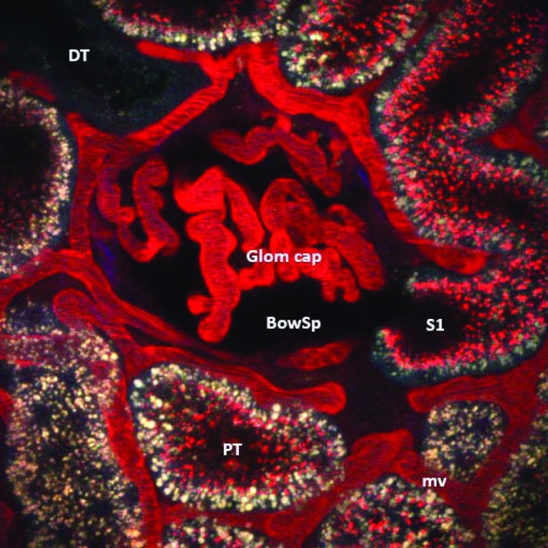
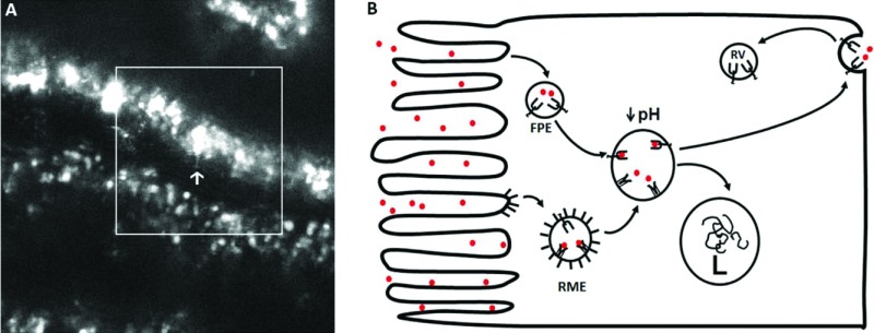
Intravital 2-photon microscopy, along with the development of fluorescent probes and innovative software, has rapidly advanced the study of intracellular and intercellular processes at the organ level. Researchers can quantify the distribution, behavior, and dynamic interactions of up to four labeled chemical probes and proteins simultaneously and repeatedly in four dimensions (3D + time) with subcellular resolution in real time. Transgenic fluorescently labeled proteins, delivery of plasmids, and photo-activatable probes enhance these possibilities. Thus, multi-photon microscopy has greatly extended our ability to understand cell biology intra-vitally at cellular and subcellular levels. For example, evaluation of rat surface glomeruli and accompanying proximal tubules has shown the long held paradigm regarding limited albumin filtration under physiologic conditions is to be questioned. Furthermore, the role of proximal tubules in determining albuminuria under physiologic and disease conditions was supported by direct visualization and quantitative analysis.
Conflict of interest statement
Potential Conflicts of Interest: None disclosed
Figures
References
-
- Dunn KW, Sandoval RM, Kelly KJ, et al. Functional studies of the kidney of living animals using multicolor two-photon microscopy. Am J Physiol Cell Physiol. 2002;283(3):C905–16. Epub 2002/08/15. doi: 10.1152/ajpcell.00159.2002. PubMed PMID: 12176747. - DOI - PubMed
-
- Hall AM, Rhodes GJ, Sandoval RM, Corridon PR, Molitoris BA. In vivo multiphoton imaging of mitochondrial structure and function during acute kidney injury. Kidney Int. 2013;83(1):72–83. doi: 10.1038/ki.2012.328. PubMed PMID: 22992467. - DOI - PMC - PubMed
-
- Molitoris BA, Sandoval RM. Intravital multiphoton microscopy of dynamic renal processes. Am J Physiol Renal Physiol. 2005;288(6):F1084–9. Epub 2005/05/11. doi: 10.1152/ajprenal.00473.2004. PubMed PMID: 15883167. - DOI - PubMed
-
- Peti-Peterdi J, Burford JL, Hackl MJ. The first decade of using multiphoton microscopy for high-power kidney imaging. Am J Physiol Renal Physiol. 2012;302(2):F227–33. doi: 10.1152/ajprenal.00561.2011. PubMed PMID: 22031850; PubMed Central PMCID: PMC3340919. - DOI - PMC - PubMed
-
- Tanner GA, Sandoval RM, Molitoris BA, Bamburg JR, Ashworth SL. Micropuncture gene delivery and intravital two-photon visualization of protein expression in rat kidney. Am J Physiol Renal Physiol. 2005;289(3):F638–43. doi: 10.1152/ajprenal.00059.2005. PubMed PMID: 15886277. - DOI - PubMed
Publication types
MeSH terms
Substances
Grants and funding
LinkOut - more resources
Full Text Sources