Primary peritoneal serous carcinoma: a rare case and palliative approach
- PMID: 25125875
- PMCID: PMC4130006
- DOI: 10.4103/0973-1075.132653
Primary peritoneal serous carcinoma: a rare case and palliative approach
Abstract
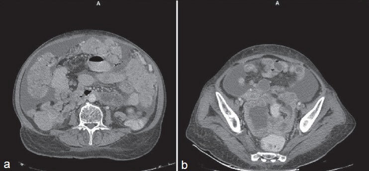
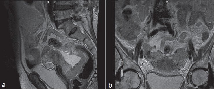
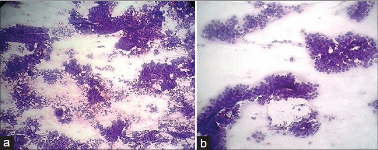
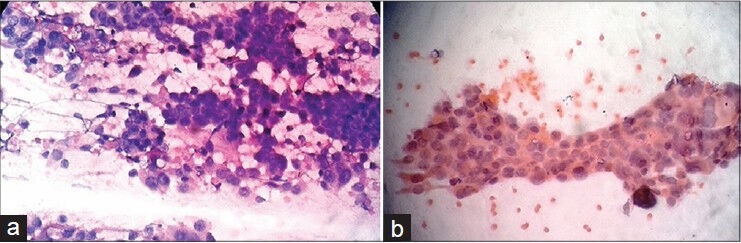
Primary peritoneal serous carcinoma (PPSC) is a rare primary malignancy that diffusely involves the peritoneum, indistinguishable clinically and histopathologically from primary serous ovarian carcinoma. The origin of PPSC has not been well characterized. Here we present a case of PPSC diagnosed in ultrasonography-guided fine needle aspiration cytology (FNAC) in a 76- old female presenting with ascites, abdominal pain, distension and constipation. PPSC is an unusual tumour but cytomorphology is distinctive enough to diagnose preoperatively. In the case report hereby described PPSC is an inoperable malignancy, hence chemotherapy and palliative care are the only offered treatment.
Keywords: Chemotherapy; Fine needle aspiration cytology; Primary peritoneal serous carcinoma.
Conflict of interest statement
Figures
Similar articles
-
Primary peritoneal serous carcinoma, an extremely rare malignancy: A case report and review of the literature.Oncol Lett. 2016 Jun;11(6):4063-4065. doi: 10.3892/ol.2016.4525. Epub 2016 May 5. Oncol Lett. 2016. PMID: 27313741 Free PMC article.
-
Primary papillary serous carcinoma of the peritoneum: report of a case with diagnosis by fine needle aspiration and immunocytochemistry.Acta Cytol. 2007 Mar-Apr;51(2):203-6. doi: 10.1159/000325717. Acta Cytol. 2007. PMID: 17425204
-
An exceptional evolution of primary peritoneal serous carcinoma.Acta Clin Belg. 2017 Dec;72(6):439-442. doi: 10.1080/17843286.2017.1300216. Epub 2017 Mar 8. Acta Clin Belg. 2017. PMID: 28271744
-
Primary peritoneal serous carcinoma: a primer for radiologists.Clin Imaging. 2022 Mar;83:56-64. doi: 10.1016/j.clinimag.2021.12.007. Epub 2021 Dec 24. Clin Imaging. 2022. PMID: 34974267 Review.
-
Peritoneal dissemination of pancreatic cancer caused by endoscopic ultrasound-guided fine needle aspiration: A case report and literature review.World J Gastroenterol. 2021 Jan 21;27(3):294-304. doi: 10.3748/wjg.v27.i3.294. World J Gastroenterol. 2021. PMID: 33519143 Free PMC article. Review.
Cited by
-
Physiotherapy Management in the Case of Primary Peritoneal Serous Carcinoma With Lower Segment Cesarean Section: A Case Report.Cureus. 2024 Feb 9;16(2):e53903. doi: 10.7759/cureus.53903. eCollection 2024 Feb. Cureus. 2024. PMID: 38465046 Free PMC article.
-
Primary peritoneal serous carcinoma, an extremely rare malignancy: A case report and review of the literature.Oncol Lett. 2016 Jun;11(6):4063-4065. doi: 10.3892/ol.2016.4525. Epub 2016 May 5. Oncol Lett. 2016. PMID: 27313741 Free PMC article.
-
Exceptional response to chemotherapy followed by concurrent radiotherapy and immunotherapy in a male with primary retroperitoneal serous Adenocarcinoma: a case report and literature review.BMC Cancer. 2019 Jul 30;19(1):748. doi: 10.1186/s12885-019-5934-4. BMC Cancer. 2019. PMID: 31362708 Free PMC article.
References
-
- Swerdlow M. Mesothelioma of the pelvic peritoneum resembling papillary cystadenocarcinoma of the ovary; case report. Am J Obstet Gynecol. 1959;77:197–200. - PubMed
-
- Muto MG, Welch WR, Mok SC, Bandera CA, Fishbaugh PM, Tsao SW, et al. Evidence for a multifocal origin of papillary serous carcinoma of the peritoneum. Cancer Res. 1995;55:490–2. - PubMed
-
- Schorge JO, Muto MG, Welch WR, Bandera CA, Rubin SC, Bell DA, et al. Molecular evidence for multifocal papillary serous carcinoma of the peritoneum in patients with germline BRCA1 mutations. J Natl Cancer Inst. 1998;80:841–5. - PubMed
Publication types
LinkOut - more resources
Full Text Sources
Other Literature Sources