Review
. 2014 Jul 30:8:132.
doi: 10.3389/fnsys.2014.00132. eCollection 2014.

Best of both worlds: promise of combining brain stimulation and brain connectome

Affiliations
Review

Best of both worlds: promise of combining brain stimulation and brain connectome

Caroline Di Bernardi Luft et al. Front Syst Neurosci. .

Abstract

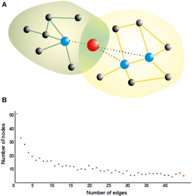
Transcranial current brain stimulation (tCS) is becoming increasingly popular as a non-pharmacological non-invasive neuromodulatory method that alters cortical excitability by applying weak electrical currents to the scalp via a pair of electrodes. Most applications of this technique have focused on enhancing motor and learning skills, as well as a therapeutic agent in neurological and psychiatric disorders. In these applications, similarly to lesion studies, tCS was used to provide a causal link between a function or behavior and a specific brain region (e.g., primary motor cortex). Nonetheless, complex cognitive functions are known to rely on functionally connected multitude of brain regions with dynamically changing patterns of information flow rather than on isolated areas, which are most commonly targeted in typical tCS experiments. In this review article, we argue in favor of combining tCS method with other neuroimaging techniques (e.g., fMRI, EEG) and by employing state-of-the-art connectivity data analysis techniques (e.g., graph theory) to obtain a deeper understanding of the underlying spatiotemporal dynamics of functional connectivity patterns and cognitive performance. Finally, we discuss the possibilities of using these combined techniques to investigate the neural correlates of human creativity and to enhance creativity.

Keywords: connectome; functional connectivity; graph theory; structural connectivity; tACS; tCS; tDCS; tRNS.


Figures

Figure 1
Figure 1
(A) A hub naturally serves as a bridge between two or more networks, as it is connected to many nodes in all of them, thereby playing an important role in brain functioning. Stimulating a hub is likely to affect many nodes even at a long distance, which could maximize the effect of stimulation. Yet networks are also vulnerable against a directed attack on the hubs, so that their inhibition, however partial and/or transient, may crucially affect the corresponding cognitive functions if it is not properly controlled (e.g., by multisite stimulation of nearby nodes). Green and orange lines: intra-network edges. Dotted lines: edges connecting the hub with nodes in both networks; (B) A typical degree distribution for brain nodes as assessed by a functional connectivity index. Most of the nodes (black) have low degree (i.e., are connected only to a few nodes); some of them (blue) have moderate degree, and a few of them (in red) are hubs, which are connected to many nodes.
Figure 2
Figure 2
Schematic representation of the possible combination of tCS and functional (EEG) brain connectivity to enhance creativity. An array of scalp electrodes, a subset of which is depicted in (A), is recorded. In (B) is possible to identify ROIs which are highly connected (yellow) and the ones that are flexible hubs (red) or less connected areas (blue) during a solution vs. non-solution RAT task. This helps identifying the target electrodes to stimulate as in (C). Stimulating these electrodes may not only eliminate the differences for the corresponding nodes, but also reduce them for areas in the same network, which are not stimulated but functionally connected to the targeted ones as depicted in (D). It is worth noting that the labels and the networks drawn in the figure are only for demonstration, as they are not precisely equivalent to their anatomical locations (they are only approximate locations on a surface, the areas in yellow are located in the medial area of the brain, which cannot be seen in a cortical mesh). The areas are abbreviated as follows: PCC, posterior cingulate cortex; PPC, posterior parietal cortex; M1, primary motor cortex; mPFC, medial prefrontal cortex; ACC, anterior cingulate cortex; DLPFC, dorsolateral prefrontal cortex; ATL, anterior temporal lobe.

References

    1. Aguirre J., Sevilla-Escoboza R., Gutiérrez R., Papo D., Buldú J. M. (2014). Synchronization of interconnected networks: the role of connector nodes. Phys. Rev. Lett. 112:248701 10.1103/PhysRevLett.112.248701 - DOI - PubMed
    1. Ali M. M., Sellers K. K., Frohlich F. (2013). Transcranial alternating current stimulation modulates large-scale cortical network activity by network resonance. J. Neurosci. 33, 11262–11275 10.1523/JNEUROSCI.5867-12.2013 - DOI - PMC - PubMed
    1. Alon G., Roys S. R., Gullapalli R. P., Greenspan J. D. (2011). Non-invasive electrical stimulation of the brain (ESB) modifies the resting-state network connectivity of the primary motor cortex: a proof of concept fMRI study. Brain Res. 1403, 37–44 10.1016/j.brainres.2011.06.013 - DOI - PubMed
    1. Amadi U., Ilie A., Johansen-Berg H., Stagg C. J. (2014). Polarity-specific effects of motor transcranial direct current stimulation on fMRI resting state networks. Neuroimage 88, 155–161 10.1016/j.neuroimage.2013.11.037 - DOI - PMC - PubMed
    1. Anderson J. R., Anderson J. F., Ferris J. L., Fincham J. M., Jung K. J. (2009). Lateral inferior prefrontal cortex and anterior cingulate cortex are engaged at different stages in the solution of insight problems. Proc. Natl. Acad. Sci. U.S.A. 106, 10799–10804 10.1073/pnas.0903953106 - DOI - PMC - PubMed