. 2014 Aug 15;9(8):e104398.
doi: 10.1371/journal.pone.0104398. eCollection 2014.

Toll-like receptor 9 mediated responses in cardiac fibroblasts

Affiliations

Toll-like receptor 9 mediated responses in cardiac fibroblasts

Ingrid Kristine Ohm et al. PLoS One. .

Abstract

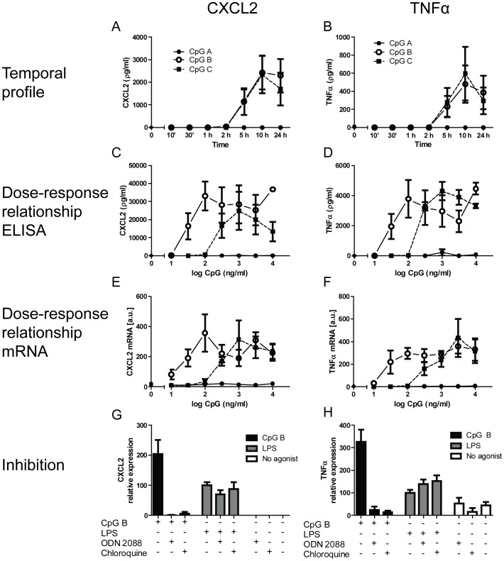
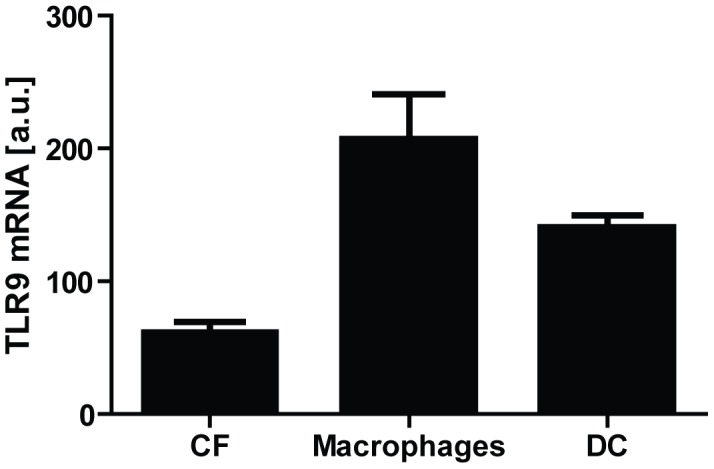
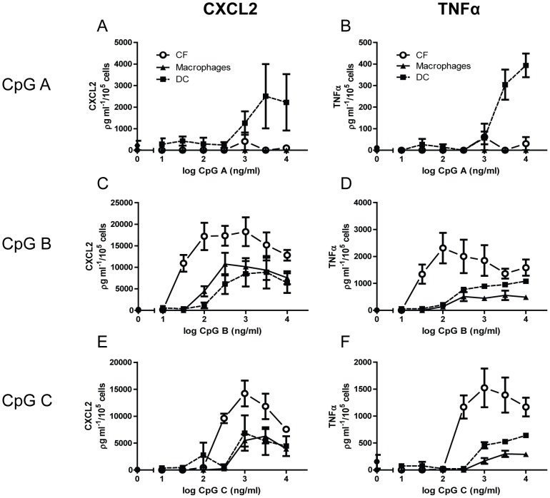
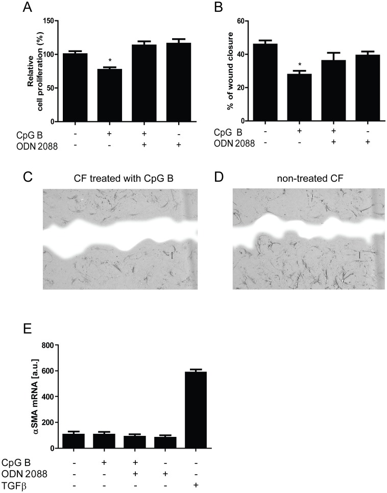
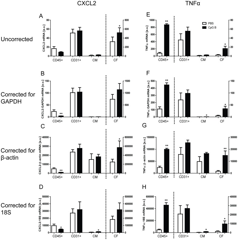
Altered cardiac Toll-like receptor 9 (TLR9) signaling is important in several experimental cardiovascular disorders. These studies have predominantly focused on cardiac myocytes or the heart as a whole. Cardiac fibroblasts have recently been attributed increasing significance in mediating inflammatory signaling. However, putative TLR9-signaling through cardiac fibroblasts remains non-investigated. Thus, our aim was to explore TLR9-signaling in cardiac fibroblasts and investigate the consequence of such receptor activity on classical cardiac fibroblast cellular functions. Cultivated murine cardiac fibroblasts were stimulated with different TLR9 agonists (CpG A, B and C) and assayed for the secretion of inflammatory cytokines (tumor necrosis factor α [TNFα], CXCL2 and interferon α/β). Expression of functional cardiac fibroblast TLR9 was proven as stimulation with CpG B and -C caused significant CXCL2 and TNFα-release. These responses were TLR9-specific as complete inhibition of receptor-stimulated responses was achieved by co-treatment with a TLR9-antagonist (ODN 2088) or chloroquine diphosphate. TLR9-stimulated responses were also found more potent in cardiac fibroblasts when compared with classical innate immune cells. Stimulation of cardiac fibroblasts TLR9 was also found to attenuate migration and proliferation, but did not influence myofibroblast differentiation in vitro. Finally, results from in vivo TLR9-stimulation with subsequent fractionation of specific cardiac cell-types (cardiac myocytes, CD45+ cells, CD31+ cells and cardiac fibroblast-enriched cell-fractions) corroborated our in vitro data and provided evidence of differentiated cell-specific cardiac responses. Thus, we conclude that cardiac fibroblast may constitute a significant TLR9 responder cell within the myocardium and, further, that such receptor activity may impact important cardiac fibroblast cellular functions.


Conflict of interest statement

Competing Interests: The authors have declared that no competing interests exist.

Figures

Figure 1
Figure 1. TLR9-stimulated responses in cardiac fibroblasts.
Temporal profiles (10 min, 30 min, 1 h, 2 h, 5 h, 10 h and 24 h) of (all at 100 ng/ml) CpG A (dots), CpG B (open circles) or CpG C (squares)-stimulated release of CXCL2 (panel A) and TNFα (panel B) in murine cardiac fibroblasts (CF). Each data point represents the mean ± SEM of 3 separate experiments. Dose-response relationships of CpG A (dots), CpG B (open circles) and CpG C (squares) –stimulated release of CXCL2 (panel C) and TNFα (panel D) at 18 hours. Each data point represents the mean ± SEM of 6 experiments. Expression levels of CXCL2 (panel E) and TNFα (panel F) were analyzed by qPCR. Each data point represents the mean ± SEM of 4 experiments. TLR9 specificity studies were performed on murine CFs exposed to CpG B (100 ng/ml) or LPS (10 ng/ml) with or without the presence of ODN 2088 (10 µg/ml) or chloroquine diphosphate (5 µg/ml). Levels of CXCL2 (panel G) and TNFα (panel H) were analyzed after 18 hours. Each data point represents the mean ± SEM of 3 experiments.
Figure 2
Figure 2. TLR9 mRNA levels in cultivated murine cardiac fibroblasts (CF; n = 3), bone marrow derived macrophages (n = 3) and dendritic cells (DC; n = 3).
Data presented as mean ± SEM.
Figure 3
Figure 3. Comparative analysis of TLR9-stimulated responses between cardiac fibroblasts and bone-marrow derived -macrophages and –dendritic cells.
Dose-response relationships of (100 ng/ml) CpG A (panels A–B), CpG B (panels C–D) and CpG C (panels E–F) stimulated release of CXCL2 (left panels) and TNFα (right panels) were examined in murine cardiac fibroblasts (CFs; circles) compared to bone marrow derived –macrophages (triangles) and –dendritic cells (DC; squares). Analysis was performed after 18 hours stimulation. Each data point represents the mean ± SEM of 3 experiments.
Figure 4
Figure 4. Functional analysis of TLR9-stimulated responses in cardiac fibroblasts.
Proliferation by BrdU-incorporation (panel A, n = 4) and proliferation/migration by scrape wound assays (panel B n = 4) were examined in murine cardiac fibroblasts (CFs). Data are presented as mean ± SEM. *p<0.05 vs. control, **p<0.01 vs. control. Panel C and D - representative photographs of CFs proliferation/migratory responses on (100 ng/ml) CpG B stimulation (panel C) and vehicle (panel D) in scrape wound assay 18 hours after cell distortion. Blue color represents wound area. Differentiation in response to CpG B (100 ng/ml) and/or ODN 2088 (1 µg/ml) was examined in murine CFs (panel E, n = 7). TGFβ (2.5 ng/ml) serves as positive control. Data are presented as mean ± SEM.
Figure 5
Figure 5. In vivo cardiac TLR9-stimulated cellular responses.
Male C57BL/6 mice were injected i.p. with 100 µl CpG B (50 µg; n = 6, black bars) or vehicle (n = 6, white bars) and euthanized after 24 hours, with subsequent isolation of cardiac myocytes (CM), CD45+, CD31+ and non-CM/non-CD45+/non-CD31+ (denominated CF). TLR9-mediated responses in the cell-fractions were analyzed by mRNA expression levels of CXCL2 (panels A–D) and TNFα (panels E–H). Uncorrected data are shown in panels A and E. CXCL2 and TNFα are also presented as corrected for GAPDH (panels B and F), β-actin (panels C and G) and 18S (panels D and H). Data presented as mean ± SEM. *p<0.05 vs. sham, **p<0.01 vs. sham.

References

    1. Medzhitov R, Janeway C Jr (2000) Innate immunity. N Engl J Med 343: 338–344. - PubMed
    1. Hemmi H, Takeuchi O, Kawai T, Kaisho T, Sato S, et al. (2000) A Toll-like receptor recognizes bacterial DNA. Nature 408: 740–745. - PubMed
    1. Krieg AM, Yi AK, Matson S, Waldschmidt TJ, Bishop GA, et al. (1995) CpG motifs in bacterial DNA trigger direct B-cell activation. Nature 374: 546–549. - PubMed
    1. Zhang Q, Raoof M, Chen Y, Sumi Y, Sursal T, et al. (2010) Circulating mitochondrial DAMPs cause inflammatory responses to injury. Nature 464: 104–107. - PMC - PubMed
    1. Oka T, Hikoso S, Yamaguchi O, Taneike M, Takeda T, et al. (2012) Mitochondrial DNA that escapes from autophagy causes inflammation and heart failure. Nature 485: 251–255. - PMC - PubMed

Publication types