. 2014 Aug 14;158(4):889-902.
doi: 10.1016/j.cell.2014.07.021.

Dissecting engineered cell types and enhancing cell fate conversion via CellNet

Affiliations

Dissecting engineered cell types and enhancing cell fate conversion via CellNet

Samantha A Morris et al. Cell. .

Abstract

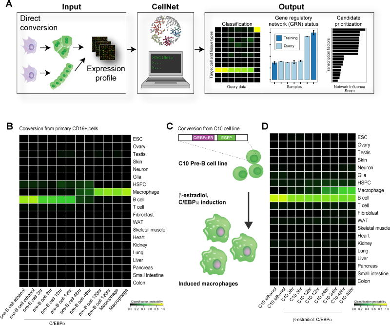
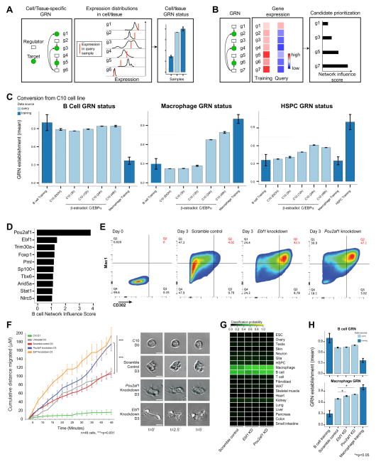
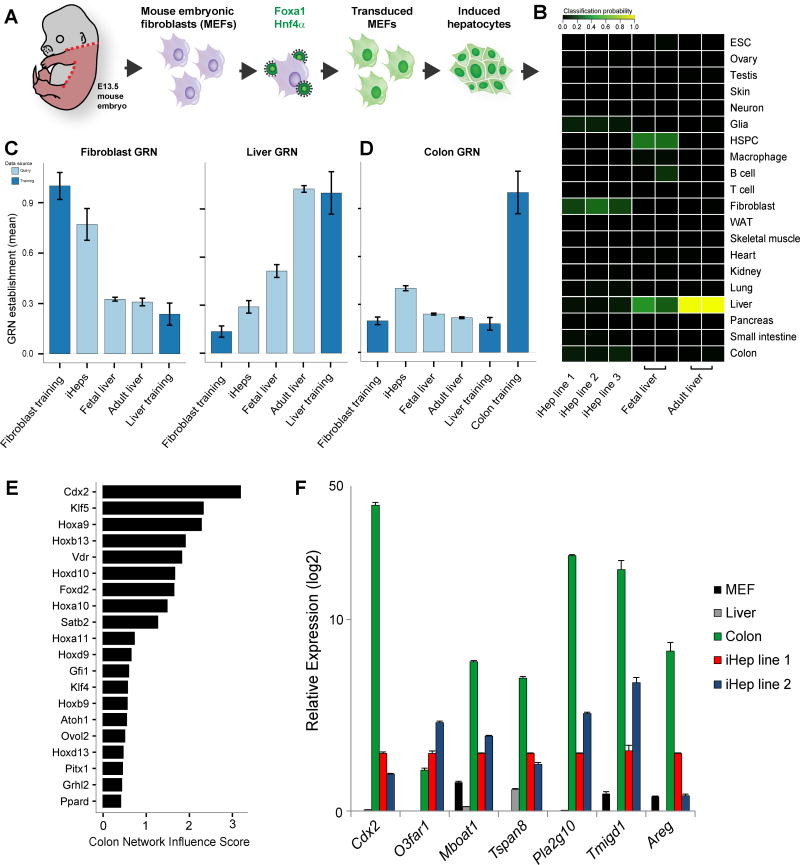
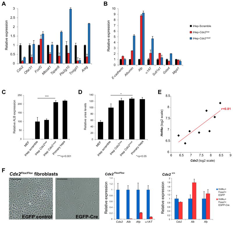
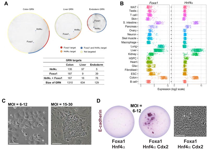
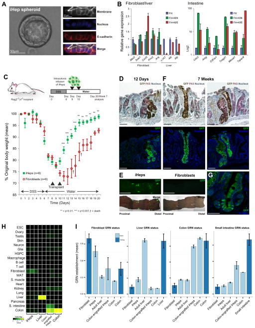
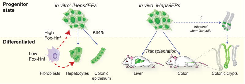
Engineering clinically relevant cells in vitro holds promise for regenerative medicine, but most protocols fail to faithfully recapitulate target cell properties. To address this, we developed CellNet, a network biology platform that determines whether engineered cells are equivalent to their target tissues, diagnoses aberrant gene regulatory networks, and prioritizes candidate transcriptional regulators to enhance engineered conversions. Using CellNet, we improved B cell to macrophage conversion, transcriptionally and functionally, by knocking down predicted B cell regulators. Analyzing conversion of fibroblasts to induced hepatocytes (iHeps), CellNet revealed an unexpected intestinal program regulated by the master regulator Cdx2. We observed long-term functional engraftment of mouse colon by iHeps, thereby establishing their broader potential as endoderm progenitors and demonstrating direct conversion of fibroblasts into intestinal epithelium. Our studies illustrate how CellNet can be employed to improve direct conversion and to uncover unappreciated properties of engineered cells.


Figures

Figure 1
Figure 1. Application of CellNet classification to the direct conversion of B cells to macrophages
(A) Adapted from Cahan et al. We designed CellNet to query gene expression profiles of engineered cell populations to classify input samples by their similarity to target cells and tissues, in order to assess the extent to which cell type and tissue GRNs are established and to score transcriptional regulators according to their likelihood of improving the engineered population. GRNs were used to construct a cell type classifier which was initially used to assess C/EBPα-mediated conversion of B cells to macrophages. (B) Each row in the classifier represents one cell or tissue type, and each column represents one array. Higher classification scores indicate a higher probability that a query sample expresses the target GRN genes at a level indistinguishable from the same cell or tissue in the training data; classification heatmap of primary B cell to macrophage conversion showing that converted cells classify exclusively as macrophages (Di Tullio et al., 2011). (C) C10 estradiol-inducible B cell to macrophage conversion (Bussmann et al., 2009). (D) Cell and tissue classification heatmap showing that conversion is not complete with induced macrophages maintaining B cell classification and only partially classifying as macrophages. See also Figure S1.
Figure 2
Figure 2. Application of the CellNet GRN status metric and candidate prioritization functions to B cell to macrophage conversion
(A) Adapted from Cahan et al. To identify cell and tissue specific GRN establishment, we devised a precise metric of GRN status. We defined this metric as the closeness of the expression of each gene in a GRN to its expected value, and then weighted each gene (g1–g6) by its importance to the network and its importance in the associated classifier. (B) To prioritize transcriptional regulator interventions, we devised a Network Influence Score integrating the target expression level of the regulator, the extent of dysregulation of the regulator and its predicted targets in the query sample, and the number of predicted targets. We applied these functions of CellNet to the estradiol-inducible B cell to macrophage conversion: (C) The B cell GRN is not extinguished and the macrophage GRN not fully established following conversion. The HPSC GRN is transiently and partially established during conversion. Data are represented as mean +/− SD. (D) CellNet prioritization of B cell transcriptional regulators whose expression is maintained in induced macrophages. (E) C/EBPα induced direct conversion of C10 B cells to macrophages following shRNA knockdown of Ebf1 and Pou2af1: flow cytometry plots of Mac1 and CD302 expression three days after conversion. The Mac1BrightCD302+ population is expanded following Ebf1 and Pou2af1 knockdown. (F) Cumulative distance migrated by induced macrophages increases following knockdowns: still images of cell morphology from timelapse imaging. Scale bars, 10μM. (G) Cell and tissue classification heatmap following knockdowns and (H) corresponding B cell and macrophage GRN establishment. *=p<0.05, t-Test. Macrophage identity is fortified following knockdown. Data are represented as mean +/− SEM. See also Figure S1, S2 and Table 1 and 2.
Figure 3
Figure 3. Application of CellNet to direct conversion of fibroblasts to hepatocytes
(A). (B) Cell and tissue classification heatmap of three independently derived iHep lines, fetal liver and adult liver: iHeps poorly classify as liver. (C) Liver and fibroblast GRN status in iHeps, fetal liver, and adult liver: the fibroblast GRN is not extinguished and liver GRN not fully established in iHeps. HSPC classification of the fetal liver reflects its role as a hematopoietic organ at this stage. (D) Partial establishment of a colon sub-GRN (424 genes) in iHeps. (E) Prioritization of transcriptional regulators of the colon GRN overexpressed in iHeps relative to native liver, led by the master intestinal regulator, Cdx2. (F) Confirmation of intestine-specific gene expression in iHeps relative to native fibroblasts, liver, and colon. Data are represented as mean +/− SEM. See also Figure S3.
Figure 4
Figure 4. Requirement of Cdx2 for function and generation of iHeps
(A) qPCR analysis of intestine-specific and liver-specific (B) gene expression following Cdx2 knockdown. Albumin expression (C) and urea production (D) in iHeps following Cdx2 knockdown. *=p<0.05, ***=p<0.001, t-Test. Cdx2 knockdown in iHeps reduces intestinal gene expression while moderately increasing liver gene expression and function, although these gains diminish with stronger Cdx2 knockdown. (E) Correlation between endogenous Hnf4α and Cdx2 expression in several iHep lines. (F) Fibroblasts fail to be converted to iHeps in the absence of Cdx2: transduction of conditional Cdx2 knockout fibroblasts. Far right: Cre expression does not impair generation of iHeps. Scale bars, 50μM. Data are represented as mean +/− SEM. See also Figure S4 and S5.
Figure 5
Figure 5. Foxa1 and Hnf4α, employed in the generation of iHeps are putative regulators of a broad endoderm GRN, as predicted by CellNet
(A) Foxa1 in particular is more heavily biased toward targets in the endoderm and colon GRNs than the liver GRN. (B) Foxa1 and Hnf4α expression over all datasets from the 20 CellNet target cells and tissues reveals their expression across a range of endoderm-derived tissues and restriction of co-expression to liver, colon, and small intestine. (C) Morphology of Foxa1-Hnf4α converted fibroblasts at MOI=6–12 (left) and MOI=15–30 (right). (D) Alkaline phosphatase staining of E-cadherin expressing iHep colonies with Foxa1-Hnf4α transduction at MOI=6–12 in the presence and absence of Cdx2. Cdx2 expression enables iHep establishment from low levels of Foxa1 and Hnf4α. Scale bars, 50μM. See also Figure S5.
Figure 6
Figure 6. iHeps are an endoderm progenitor harboring intestinal identity and can functionally engraft mouse colon
(A) Culture of iHeps as spheroids and determination of cell polarity by immunofluorescence staining of E-cadherin. (B) qPCR analysis of fibroblast, liver and intestinal gene expression following three-factor driven conversion to ‘Klf-iEPs’ with Hnf4α, Foxa1 and Klf4 or Klf5 (C) Functional engraftment of GFP-expressing iHeps into mice with DSS-induced colitis. Weight gain following DSS withdrawal in GFP-iHep/iEP recipient animals: the latter regain weight significantly faster than recipients of GFP-fibroblasts, **=p<0.01, ***=p<0.001, t-Test. (D) Immunohistochemistry and immunofluorescence of short-term engrafted GFP-iHeps/iEPs in serial sections of colon 12 days after transplantation. (E) Whole-mounts of engrafted GFP-iHeps/iEPs. Engraftment of GFP-fibroblasts is not observed. Scale bars, 0.5cm. (F) Long-term engrafted GFP-iHeps/iEPs in the colon 7 weeks after transplantation. Goblet cells are visualized by PAS staining. Scale bars, 100μm. (G) Detail of long-term engrafted crypt. (H) CellNet classification of engrafted iHeps/iEPs recovered 12 days following transplantation. Colon classification is strongly fortified in engrafted iHeps/iEPs, comparable to native colon, relative to in vitro cultured iHeps/iEPs. (I) Fibroblast, liver and broad colon GRN status in colon-engrafted iHeps/iEPs: Fibroblast and liver GRNs are extinguished where colon GRN status is fortified following maturation in vivo. Data are represented as mean +/− SEM. Scale bars as indicated. See also Figure S6.
Figure 7
Figure 7. Model: Foxa1 and Hnf4α specify an endoderm progenitor state
Left: In this study we find that low levels of Foxa1 and Hnf4α expression in fibroblasts in vitro specifies cells bearing the hallmarks of differentiated hepatocytes. High levels of Foxa1 and Hnf4α specifies a progenitor state, ‘iHeps/iEPs’ from which differentiation toward an intestinal fate, ‘Klf-iEPs’, can be coaxed by Klf4/5 expression. Right: Transplantation of iHeps/iEPs into mouse liver (as in Sekiya and Suzuki, 2011) or mouse colon leads to differentiation of progenitors to support long-term engraftment of the colon suggesting that iHeps/iEPs possess intestinal stem-like properties.

Comment in

References

    1. Barker N, van Es JH, Kuipers J, Kujala P, van den Born M, Cozijnsen M, Haegebarth A, Korving J, Begthel H, Peters PJ, et al. Identification of stem cells in small intestine and colon by marker gene Lgr5. Nature. 2007;449:1003–1007. - PubMed
    1. Bernardo GM, Keri RA. FOXA1: a transcription factor with parallel functions in development and cancer. Biosci Rep. 2012;32:113–130. - PMC - PubMed
    1. Bussmann LH, Schubert A, Vu Manh TP, De Andres L, Desbordes SC, Parra M, Zimmermann T, Rapino F, Rodriguez-Ubreva J, Ballestar E, et al. A robust and highly efficient immune cell reprogramming system. Cell Stem Cell. 2009;5:554–566. - PubMed
    1. Cahan P, Li H, Morris SA, Lummertz da Rocha E, Daley GQ, Collins JJ. CellNet: Network Biology Applied to Stem Cell Engineering - PMC - PubMed
    1. Cohen DE, Melton D. Turning straw into gold: directing cell fate for regenerative medicine. Nat Rev Genet. 2011;12:243–252. - PubMed

Publication types

Associated data