. 2014 Aug 15;9(8):e104596.
doi: 10.1371/journal.pone.0104596. eCollection 2014.

The X gene of adeno-associated virus 2 (AAV2) is involved in viral DNA replication

Affiliations

The X gene of adeno-associated virus 2 (AAV2) is involved in viral DNA replication

Maohua Cao et al. PLoS One. .

Abstract

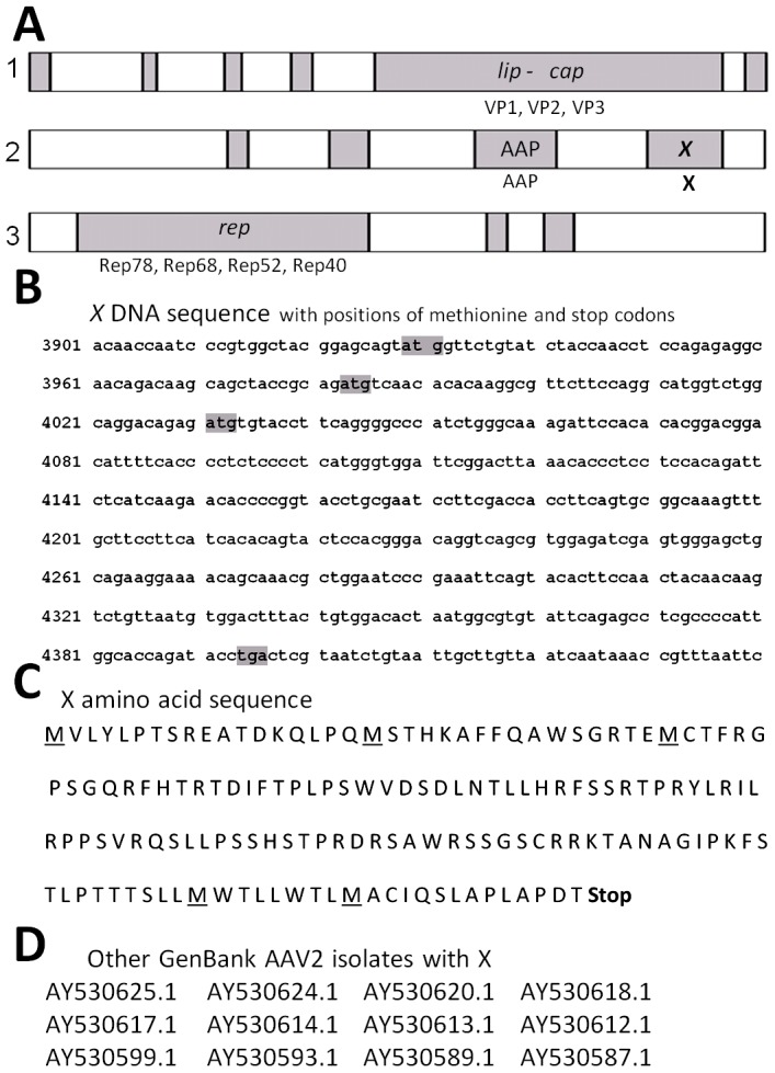
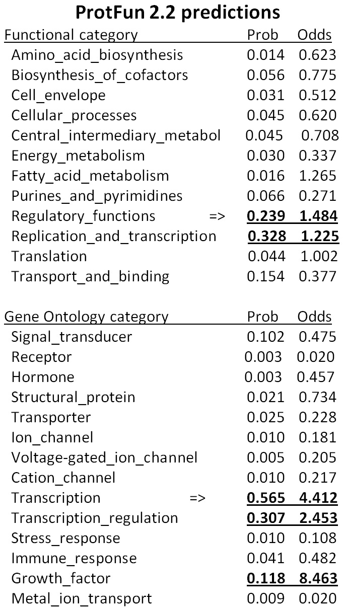
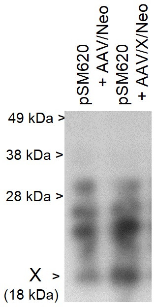
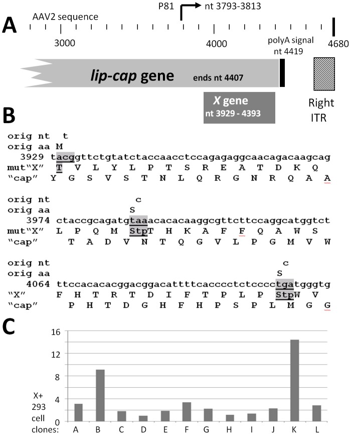
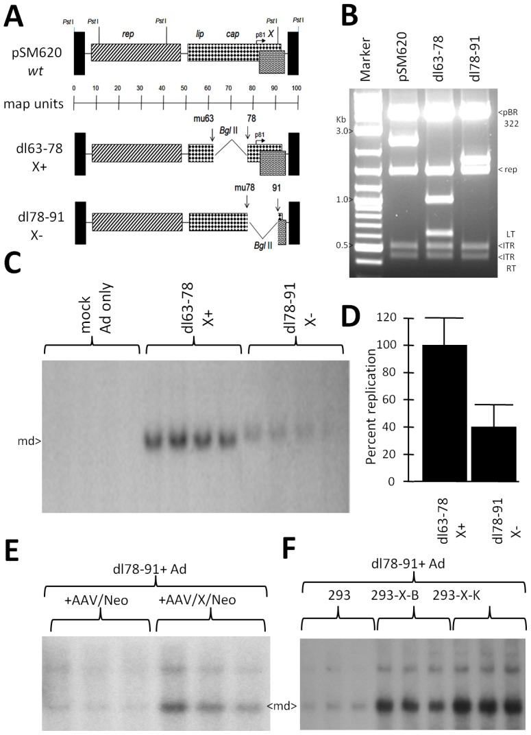
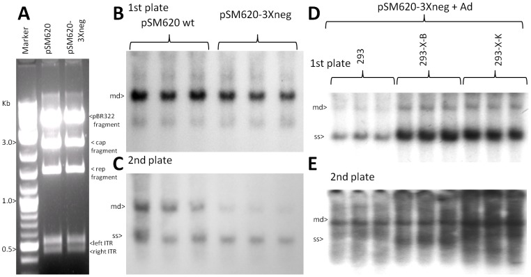
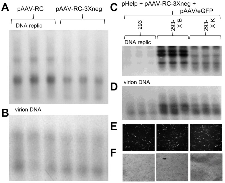
Adeno-associated virus (AAV) (type 2) is a popular human gene therapy vector with a long active transgene expression period and no reported vector-induced adverse reactions. Yet the basic molecular biology of this virus has not been fully addressed. One potential gene at the far 3' end of the AAV2 genome, previously referred to as X (nt 3929 to 4393), overlapping the 3' end of the cap gene, has never been characterized, although we did previously identify a promoter just up-stream (p81). Computer analysis suggested that X was involved in replication and transcription. The X protein was identified during active AAV2 replication using a polyclonal antibody against a peptide starting at amino acid 98. Reagents for the study of X included an AAV2 deletion mutant (dl78-91), a triple nucleotide substitution mutant that destroys all three 5' AUG-initiation products of X, with no effect on the cap coding sequence, and X-positive-293 cell lines. Here, we found that X up-regulated AAV2 DNA replication in differentiating keratinocytes (without helper virus, autonomous replication) and in various forms of 293 cell-based assays with help from wild type adenovirus type 5 (wt Ad5) or Ad5 helper plasmid (pHelper). The strongest contribution by X was seen in increasing wt AAV2 DNA replication in keratinocytes and dl78-91 in Ad5-infected X-positive-293 cell lines (both having multi-fold effects). Mutating the X gene in pAAV-RC (pAAV-RC-3Xneg) yielded approximately a ∼33% reduction in recombinant AAV vector DNA replication and virion production, but a larger effect was seen when using this same X-knockout AAV helper plasmid in X-positive-293 cell lines versus normal 293 cells (again, multi-fold). Taken together these data strongly suggest that AAV2 X encodes a protein involved in the AAV life cycle, particularly in increasing AAV2 DNA replication, and suggests that further studies are warranted.


Conflict of interest statement

Competing Interests: The authors have declared that no competing interests exist.

Figures

Figure 1
Figure 1. Sequences of AAV2 X.
A shows the open reading frames (ORF), reading from the natural AAV2 promoters (left to right), as analyzed by NIH ORF finder software analysis of NC_001401 (AAV2, Kleinschmidt) with their names/functions indicated at the top of the figure as determined by mutational analysis . B shows the nucleotide (nt) sequence of the third largest ORF, called X , of AAV2 with its start methionines and stop codon highlighted in grey. C shows the amino acid (aa) sequence of the X. D shows a series of AAV2 isolates found in Genbank which also show the X ORF.
Figure 2
Figure 2. Software predicted functions of X.
A shown are results from Technical University of Denmark, ProtFun 2.2 software analysis which attempts to predict functions of proteins. Notice that replication and transcription are listed multiple times.
Figure 3
Figure 3. Identification of X protein.
Shown is a Western blot of protein from 293 cells infected with Ad5 and transfected with pSM620 (wt AAV2) plus either AAV/Neo or AAV/X/Neo. The Western blot was probed with polyclonal rabbit antibodies directed against a peptide derived from aa 98–111of AAV2 X. While polyclonal antibodies are well known for having cross-reactivity, note that a protein of approximately 18 kDa, the predicted size of X, is seen strongly enhanced in cells transfected with AAV/X/Neo, consistent with X.
Figure 4
Figure 4. Environs of the X gene and reagents for X study.
A shows the region of X at the 3′ end of AAV2. Included are the 3′ end of lip-cap , , the p81 promoter , the poly A sequence and the 3′, right, inverted terminal repeat (ITR). B shows three nt substitution mutations in X which eliminate the products from all three 5′/amino end X start methionines, but which have no effect on the cap ORF/coding sequence. C shows the analysis of twelve 293 cell clones generated by transfection of pCI/X/Neo, and then G418 selected. The left scale shows the copy number of X found by Q-RT-PCR with clone D as the “1X” reference clone. 293-X-B and 293-X-K, having the highest copy numbers of X were chosen for further study.
Figure 5
Figure 5. X enhances AAV2 autonomous DNA replication in skin rafts.
A shows the structure of the AAV vector plasmids used. B shows the structure of the experiment analyzing X gene function in the skin raft (stratified squamous epithelium, autonomous AAV2 replication). Note that the plasmid is transfected before infection of the keratinocytes with wt AAV. This is done so as to allow the transfected gene to be expressed during the early phase of wt AAV replication. C shows the resulting Southern blot of DNA after probing the membrane with 32P-cap sequences (but not including X sequences). D shows a quantification of five such experiments. Note that AAV2 DNA replication is enhanced 6 fold. E shows that X enhances AAV2 rep expression relative to housekeeping TFIIB gene expression. These data are fully consistent with the higher DNA replication found in C. F shows dosage dependent affect of adding X. Note that the larger the amount of AAV/X/Neo transfected the higher the level of AAV2 DNA replication.
Figure 6
Figure 6. Deletion of X gives lower DNA replication of AAV2.
A shows the structure of AAV2 deletion mutants dl63-78 and dl78-91, with wild type (wt) AAV2 shown at the top, including Pst I restriction sites. B shows a Pst I, Bgl II dual digestion of dl63-78 and dl78-91. C shows a Southern blot analysis comparison of dl63-78 and dl78-91 DNA replication in Ad5-infected 293 cells, probed with 32P-rep DNA, and densitometrically quantitated in D. Note that dl63-78 replicates approximately 2.5 fold higher than the dl78-91(p<0.05). E shows a comparison of dl78-91 DNA replication upon co-transfection with either AAV/Neo or AAV/X/Neo into Ad5-infected 293 cells (p<0.05). F shows a Southern blot analysis comparison of dl78-91 DNA replication in Ad5-infected unaltered 293 cells, 293-X-B, and 293-X-K, probed with 32P-rep DNA. An analysis of the level of copy numbers of X in these cells is shown in Figure 4 C. Note that dl78-91 replicates to higher levels in the 293 cells which contain the X gene (complementation) compared to unaltered 293 cells without X (p<0.05).
Figure 7
Figure 7. pSM620-3Xneg, without X, displays weaker DNA replication in Ad5-infected 293 cells.
A shows a Pst I restriction digestion analysis of wt pSM620 and pSM620-3Xneg (X-). B shows the Southern blot of DNA replication, using 32P-rep pribe, of pSM620 and pSM620-3Xneg relative to each other in Ad5-infected 293 cells. Note that pSM620 replicated to a slightly higher level than pSM620-3Xneg (p = 0.057). C shows a “2nd plate analysis” where equal aliquots of virus stock from plates identical to those of B where heated to 56°C (to kill Ad5), and then used to infect a second plate of Ad5-infected 293 cells. Shown is the Southern blot of DNA replication, using P32-rep probe, of pSM620 and pSM620-3Xneg replication from resulting first plate generated virus infection (p<0.05). D shows a Southern blot analysis comparison of pSM620-3Xneg replication in Ad5-infected unaltered 293 cells, 293-X-B, and 293-X-K, probed with 32P-rep DNA. Note that pSM620-3Xneg replicates to higher levels in the 293 cells which contain the X gene (complementation) compared to 293 cells without X (p<0.05). E shows another “2nd plate analysis” where equal aliquots of virus stock from plates identical to those of D, which where heated to 56°C (to kill Ad5), and then used to infect a second plate of Ad5-infected 293 (normal) cells. Shown is the Southern blot of DNA replication, using 32P-rep probe, of pSM620-3Xneg replication from resulting first plate generated virus infection. Note that, pSM620-3Xneg replicated to higher levels in the 2nd plate (p<0.05) due to higher levels of virus produced in the first plate.
Figure 8
Figure 8. Recombinant defective (r)AAV DNA replication and virion production are lower without X.
A shows the Southern blot analysis of rAAV/eGFP DNA replication, using 32P-eGFP probe, resulting from the standard 293 cell triple transfection procedure (pAAV/eGFP, pHelper, pAAV-RC) except comparing the usage of either wt pAAV-RC or pAAV-RC-3Xneg. Note that use of pAAV-RC resulted in slightly higher pAAV/eGFP DNA replication levels than when using pAAV-RC-3Xneg (p<0.05). B shows a Southern blot analysis of DNAse I resistant virion DNA (encapsidated genomes). Again note that the use of wt pAAV-RC resulted in higher rAAV/eGFP virion levels (p<0.05). C shows a Southern blot which (32P-eGFP probe) which compares the use of pAAV-RC-3Xneg, along with pAAV/eGFP and pHelp, to replicate AAV/eGFP DNA in unaltered 293, versus 293-X-B and 293-X-K cells, both of which contain the X gene. Note that higher DNA replication levels of AAV/eGFP take place in the X-positive 293-X-B and 293-X-K cells than normal 293 cells (p<0.05). D shows a Southern blot analysis of DNAse I resistant virion DNA (encapsidated genomes). Again note that the use of 293-X-B and 293-X-K cells, having the X gene, resulted in higher rAAV/eGFP virion levels (p<0.05). E shows an analysis of eGFP expression/virion infectivity in which AAV/eGFP virus, equalized for comparable titers from quantitative densitometric analysis of the virion DNA Southern blot in panel D was used to infect normal 293 cells and analyzed for eGFP expression at two days post-infection. Note that equal eGFP expression can be seen across all three cell infections indicating that the use of pAAV-RC-3Xneg with the 293-X-positive cell lines gave virus with comparable infectivity to the standard pAAV-RC/wt 293 cell production scheme. F show a white light picture of the same field depicted in E as a control for cell viability.

References

    1. Hermonat PL, Labow MA, Wright R, Berns KI, Muzyczka N (1984) Genetics of adeno-associated virus: isolation and preliminary characterization of mutants in adeno-associated virus type 2. J Virol 51: 329–339. - PMC - PubMed
    1. Hermonat PL, Muzyczka N (1984) Use of adeno-associated virus as a mammalian DNA cloning vector: transduction of neomycin resistance into mammalian tissue culture cells. Proc Natl Acad Sci USA 81: 6466–6470. - PMC - PubMed
    1. Hermonat PL (1984) Genetic analysis and utilization of adeno-associated virus as a mammalian cloning vector. Univ FL Dissertation, Available: http://archive.org/details/geneticanalysisu00herm.
    1. Hermonat PL (2014) The first adeno-associated virus gene transfer experiment, 1983. In press Human Gene Therapy - PubMed
    1. You CX, Shi M, Liu Y, Cao M, Luo RC, et al. (2012) AAV2/IL-12 gene delivery into dendritic cells (DC) enhances CTL stimulation above other IL-12 applications: evidence for IL-12 intracrine activity in DC. Oncoimmunology 1: 847–855. - PMC - PubMed

Publication types