Therapeutic potential of a non-steroidal bifunctional anti-inflammatory and anti-cholinergic agent against skin injury induced by sulfur mustard
- PMID: 25127551
- PMCID: PMC4254337
- DOI: 10.1016/j.taap.2014.07.016
Therapeutic potential of a non-steroidal bifunctional anti-inflammatory and anti-cholinergic agent against skin injury induced by sulfur mustard
Abstract
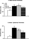
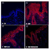
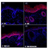
Sulfur mustard (bis(2-chloroethyl) sulfide, SM) is a highly reactive bifunctional alkylating agent inducing edema, inflammation, and the formation of fluid-filled blisters in the skin. Medical countermeasures against SM-induced cutaneous injury have yet to be established. In the present studies, we tested a novel, bifunctional anti-inflammatory prodrug (NDH 4338) designed to target cyclooxygenase 2 (COX2), an enzyme that generates inflammatory eicosanoids, and acetylcholinesterase, an enzyme mediating activation of cholinergic inflammatory pathways in a model of SM-induced skin injury. Adult SKH-1 hairless male mice were exposed to SM using a dorsal skin vapor cup model. NDH 4338 was applied topically to the skin 24, 48, and 72 h post-SM exposure. After 96 h, SM was found to induce skin injury characterized by edema, epidermal hyperplasia, loss of the differentiation marker, keratin 10 (K10), upregulation of the skin wound marker keratin 6 (K6), disruption of the basement membrane anchoring protein laminin 322, and increased expression of epidermal COX2. NDH 4338 post-treatment reduced SM-induced dermal edema and enhanced skin re-epithelialization. This was associated with a reduction in COX2 expression, increased K10 expression in the suprabasal epidermis, and reduced expression of K6. NDH 4338 also restored basement membrane integrity, as evidenced by continuous expression of laminin 332 at the dermal-epidermal junction. Taken together, these data indicate that a bifunctional anti-inflammatory prodrug stimulates repair of SM induced skin injury and may be useful as a medical countermeasure.
Keywords: Non-steroid bifunctional anti-inflammatory and anti-cholinergic agent; Skin; Sulfur mustard; Wound repair.
Copyright © 2014 Elsevier Inc. All rights reserved.
Conflict of interest statement
The authors declare that there are no conflicts of interest.
Figures


Similar articles
-
Tissue injury and repair following cutaneous exposure of mice to sulfur mustard.Ann N Y Acad Sci. 2016 Aug;1378(1):118-123. doi: 10.1111/nyas.13125. Epub 2016 Jul 2. Ann N Y Acad Sci. 2016. PMID: 27371823 Free PMC article. Review.
-
Sulfur mustard induced mast cell degranulation in mouse skin is inhibited by a novel anti-inflammatory and anticholinergic bifunctional prodrug.Toxicol Lett. 2018 Sep 1;293:77-81. doi: 10.1016/j.toxlet.2017.11.005. Epub 2017 Nov 7. Toxicol Lett. 2018. PMID: 29127031 Free PMC article.
-
Structural changes in the skin of hairless mice following exposure to sulfur mustard correlate with inflammation and DNA damage.Exp Mol Pathol. 2011 Oct;91(2):515-27. doi: 10.1016/j.yexmp.2011.05.010. Epub 2011 Jun 13. Exp Mol Pathol. 2011. PMID: 21672537 Free PMC article.
-
Time course of skin features and inflammatory biomarkers after liquid sulfur mustard exposure in SKH-1 hairless mice.Toxicol Lett. 2015 Jan 5;232(1):68-78. doi: 10.1016/j.toxlet.2014.09.022. Epub 2014 Sep 30. Toxicol Lett. 2015. PMID: 25275893
-
Skin Models Used to Define Mechanisms of Action of Sulfur Mustard.Disaster Med Public Health Prep. 2023 Oct 18;17:e551. doi: 10.1017/dmp.2023.177. Disaster Med Public Health Prep. 2023. PMID: 37849329 Free PMC article. Review.
Cited by
-
Expression of cytokines and chemokines in mouse skin treated with sulfur mustard.Toxicol Appl Pharmacol. 2018 Sep 15;355:52-59. doi: 10.1016/j.taap.2018.06.008. Epub 2018 Jun 20. Toxicol Appl Pharmacol. 2018. PMID: 29935281 Free PMC article.
-
Defining the timing of 25(OH)D rescue following nitrogen mustard exposure.Cutan Ocul Toxicol. 2018 Jun;37(2):127-132. doi: 10.1080/15569527.2017.1355315. Epub 2017 Aug 8. Cutan Ocul Toxicol. 2018. PMID: 28737434 Free PMC article.
-
A standardized in vitro bioengineered skin for penetrating wound modeling.In Vitro Model. 2025 Feb 21;4(1):15-30. doi: 10.1007/s44164-025-00082-x. eCollection 2025 Feb. In Vitro Model. 2025. PMID: 40160210 Free PMC article.
-
Tissue injury and repair following cutaneous exposure of mice to sulfur mustard.Ann N Y Acad Sci. 2016 Aug;1378(1):118-123. doi: 10.1111/nyas.13125. Epub 2016 Jul 2. Ann N Y Acad Sci. 2016. PMID: 27371823 Free PMC article. Review.
-
Scar management in burn injuries using drug delivery and molecular signaling: Current treatments and future directions.Adv Drug Deliv Rev. 2018 Jan 1;123:135-154. doi: 10.1016/j.addr.2017.07.017. Epub 2017 Jul 27. Adv Drug Deliv Rev. 2018. PMID: 28757325 Free PMC article. Review.
References
-
- Amano S. Possible involvement of basement membrane damage in skin photoaging. J Investig Dermatol Symp Proc. 2009;14:2–7. - PubMed
-
- Babin MC, Ricketts K, Skvorak JP, Gazaway M, Mitcheltree LW, Casillas RP. Systemic administration of candidate antivesicants to protect against topically applied sulfur mustard in the mouse ear vesicant model (MEVM) J Appl Toxicol. 2000;20(Suppl 1):S141–S144. - PubMed
-
- Black AT, Joseph LB, Casillas RP, Heck DE, Gerecke DR, Sinko PJ, Laskin DL, Laskin JD. Role of MAP kinases in regulating expression of antioxidants and inflammatory mediators in mouse keratinocytes following exposure to the half mustard, 2-chloroethyl ethyl sulfide. Toxicol Appl Pharmacol. 2010;245:352–360. - PMC - PubMed
-
- Casillas RP, Kiser RC, Truxall JA, Singer AW, Shumaker SM, Niemuth NA, Ricketts KM, Mitcheltree LW, Castrejon LR, Blank JA. Therapeutic approaches to dermatotoxicity by sulfur mustard. I. Modulaton of sulfur mustard-induced cutaneous injury in the mouse ear vesicant model. J Appl Toxicol. 2000;20(Suppl 1):S145–S151. - PubMed
Publication types
MeSH terms
Substances
Grants and funding
LinkOut - more resources
Full Text Sources
Other Literature Sources
Medical
Molecular Biology Databases
Research Materials
Miscellaneous

