. 2014 Oct 15;394(2):327-39.
doi: 10.1016/j.ydbio.2014.07.024. Epub 2014 Aug 13.

In vivo analysis of hyaloid vasculature morphogenesis in zebrafish: A role for the lens in maturation and maintenance of the hyaloid

Affiliations

In vivo analysis of hyaloid vasculature morphogenesis in zebrafish: A role for the lens in maturation and maintenance of the hyaloid

Andrea Hartsock et al. Dev Biol. .

Abstract

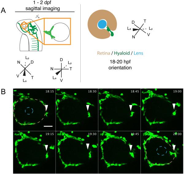
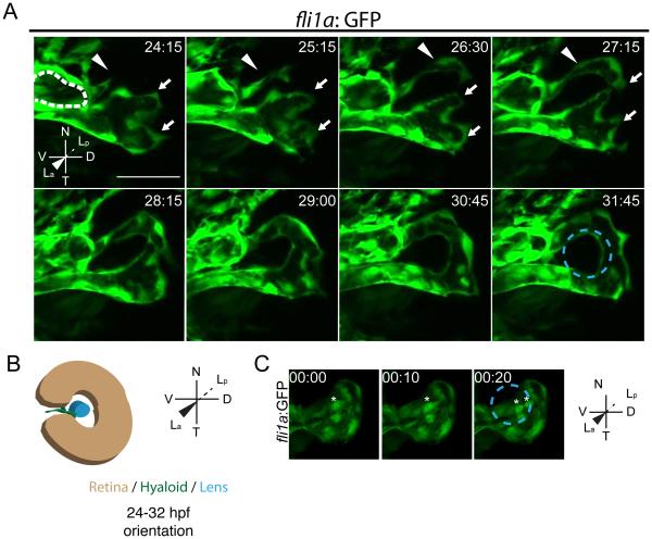
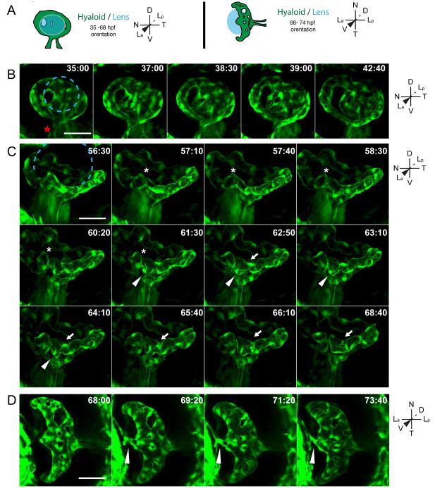
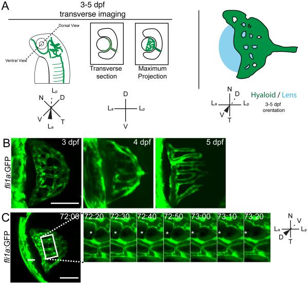
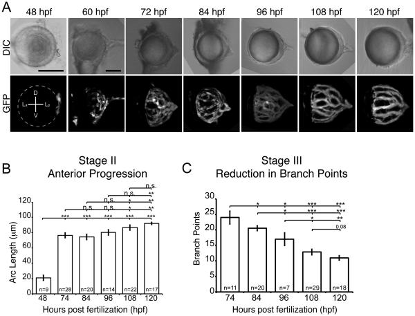
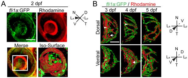
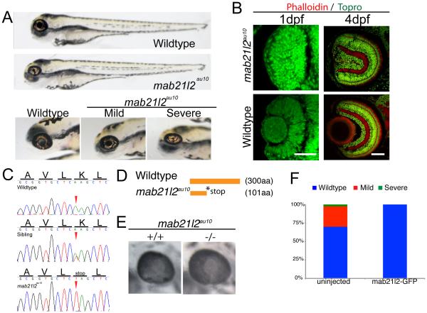
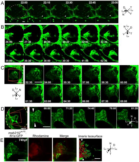
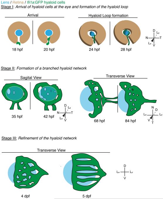
Two vascular networks nourish the embryonic eye as it develops - the hyaloid vasculature, located at the anterior of the eye between the retina and lens, and the choroidal vasculature, located at the posterior of the eye, surrounding the optic cup. Little is known about hyaloid development and morphogenesis, however. To begin to identify the morphogenetic underpinnings of hyaloid formation, we utilized in vivo time-lapse confocal imaging to characterize morphogenesis of the zebrafish hyaloid through 5 days post fertilization (dpf). Our data segregate hyaloid formation into three distinct morphogenetic stages: Stage I: arrival of hyaloid cells at the lens and formation of the hyaloid loop; Stage II: formation of a branched hyaloid network; Stage III: refinement of the hyaloid network. Utilizing fixed and dissected tissues, distinct Stage II and Stage III aspects of hyaloid formation were quantified over time. Combining in vivo imaging with microangiography, we demonstrate that the hyaloid system becomes fully enclosed by 5dpf. To begin to identify the molecular and cellular mechanisms underlying hyaloid morphogenesis, we identified a recessive mutation in the mab21l2 gene, and in a subset of mab21l2 mutants the lens does not form. Utilizing these "lens-less" mutants, we determined whether the lens was required for hyaloid morphogenesis. Our data demonstrate that the lens is not required for Stage I of hyaloid formation; however, Stages II and III of hyaloid formation are disrupted in the absence of a lens, supporting a role for the lens in hyaloid maturation and maintenance. Taken together, this study provides a foundation on which the cellular, molecular and embryologic mechanisms underlying hyaloid morphogenesis can be elucidated.

Keywords: Eye development; Hyaloid vasculature; Lens; Zebrafish.


Figures

Fig. 1
Fig. 1
Stage I: Arrival of fli1a:GFP+ cells at the ventral eye. (A) Schematic of 1-2dpf imaging paradigm and maximum projection data. Drawings not to scale. (B) fli1a:GFP+ cells (arrowheads) arrive at the ventral eye between 18-20hpf. Precursor cells proceed towards the lens through the choroid fissure. Lens position indicated by dashed blue line, retina by dashed beige line. hh:mm. Scale bar = 50μm D:Dorsal, V:Ventral, N:Nasal, T:Temporal, La:Lens anterior, Lp:Lens posterior
Fig. 2
Fig. 2
Stage I: Hyaloid loop formation. (A) Still maximum projection images from time-lapse movies highlighting formation of the hyaloid loop. Nasal (white arrow) and temporal (white arrowhead) oriented sprout morphogenesis to form the hyaloid loop. Dashed white line outlines cross section of vessel from which the hyaloid loop sprouts. (B) Schematic of maximum projection data. (C) Stills from time-lapse movie showing mitotic cells in the vessel stalk (asterisks) during formation of hyaloid loop. Lens position in B and C indicated by dashed blue line. hh:mm. All scale bars = 50μm. D:Dorsal, V:Ventral, N:Nasal, T:Temporal, La:Lens anterior, Lp:Lens posterior.
Fig. 3
Fig. 3
Stage II: Formation of a branched hyaloid network. All images are still maximum projections from time-lapse movies. (A) Schematic of imaging angles and maximum projection data. Drawings not to scale. (B) Increases in vessel network complexity within the posterior hyaloid. Asterisk marks the nasally oriented sprout of the hyaloid loop. (C) Angiogenesis within the hyaloid increases vessel network complexity. Asterisk marks site of break in vessel, arrowheads mark sprouting event that does not form a new connection, arrows mark sprouting event that generates a new loop within the vessel network. (D) Connection of the hyaloid to the annular ring (arrowhead). Lens position in B and C indicated by dashed blue line. hh:mm. Scale bars = 50μm. D:Dorsal, V:Ventral, N:Nasal, T:Temporal, La:Lens anterior, Lp:Lens posterior.
Fig. 4
Fig. 4
Stage III: Refinement of the hyaloid network. (A) Schematic of imaging angles and maximum projections data. Drawings not to scale. (B) Maximum projections of 3-5dpf hyaloid in transverse imaging view demonstrating the continued reduction and refinement of the vessel network. (C) Still images from time-lapse movies demonstrating the further reduction in vessel complexity as fibers connecting adjacent vessels retract (asterisk). hh:mm. (A,B) Scale bar = 50μm; inset scale bar = 5μm. D:Dorsal, V:Ventral, N:Nasal, T:Temporal, La:Lens anterior, Lp:Lens posterior.
Fig. 5
Fig. 5
Quantification of hyaloid growth. (A) DIC and GFP maximum projections images of dissected lenses from fli1a:GFP embryos at 12 hour intervals from 2-5dpf. Dashed line indicates location of the lens. Scale bars = 50 μm. D:Dorsal, V:Ventral, La:Lens anterior, Lp:Lens posterior (B) Quantification of anterior progression of the hyaloid over time. (C) Reduction in hyaloid vessel branching over time. * p ≥ 0.01 , ** p ≥ 0.001, *** p ≥ 1x10⁻¹⁰. n = number of samples analyzed.
Fig. 6
Fig. 6
Microangiography demonstrates that the hyaloid vessel is fully enclosed by 5dpf. (A) Maximum projection images of the eye of a fli1a:GFP (green) embryo injected with 50 kDa rhodamine dextran (red). Merged image highlights that rhodamine is detected throughout the vitreous. Iso-surface rendering of merged image. (B-D) Iso-surface renderings of maximum projections collected from the eyes of 3-5dpf fli1a:GFP (green) embryos injected with 50 kDa rhodamine dextran (red) and imaged either dorsally or ventrally. Arrowhead highlights rhodamine containment in a fli1a:GFP vessel and arrows highlight rhodamine outside of vessels. hh:mm. Scale bars = 50μm. D:Dorsal, V:Ventral, N:Nasal, T:Temporal, La:Lens anterior, Lp:Lens posterior.
Fig. 7
Fig. 7
mab21l2au10 mutants possess defects in lens formation. (A) Images of phenotypically wild-type sibling and mab21l2au10 mutants at 4dpf. High-magnification views of the eyes of mild and severe mab21l2 au10 mutants. (B) Transverse cryossections of wild-type and severe mab21l2au10 mutants at 1 and 4dpf highlighting the lack of a lens in severe mab21l2au10 mutants. Scale bars = 50 μm. (C) Genomic sequences from wild-type, heterozygous and mab21l2au10 mutants. mab21l2au10 mutants possess a an A→T transversion at position 301, resulting in a premature stop codon at amino acid 101. (D) Schematic of protein length of wild-type and mab21l2au10 mutant. (E) High magnification view of eye in sibling (wild-type) and mab21l2au10 mutant embryo injected with mab21l2–GFP (rescue). (F) Quantification of lens phenotype after mab21l2–GFP injection.
Fig. 8
Fig. 8
The lens is required for Stage II and III hyaloid maturation and maintenance. All images are stills from time-lapse movies from severe mab21l2au10;fli1a:GFP mutants. (A) Hyaloid precursor cell recruitment is delayed by ~2 hours in mab21l2au10 mutants but recruitment is otherwise normal. (B) Hyaloid loop formation occurs in the absence of a lens. (C) Stage II formation of a branched hyaloid network is disrupted in mab21l2au10 mutants. Hyaloid cells appear disorganized and dynamic, having not coalesced into obvious vessels. (D) The hyaloid in mab21l2au10 mutants still makes contact anteriorly with the annular vessel. (E) Microangiography demonstrates that at 3dpf, the mab21l2au10 mutant hyaloid is not enclosed anteriorly, and rhodamine (red) fills the retina. hh:mm. Beige dashed line in A, B and D indicate outlines of the retina. All scale bars = 50μm. D:Dorsal, V:Ventral, N:Nasal, T:Temporal, La:Lens anterior, Lp:Lens posterior.
Fig. 9
Fig. 9
Schematic depiction of hyaloid morphogenesis in zebrafish. See text for details of each stage. Drawings are not to scale.

References

    1. Adams RH, Alitalo K. Molecular regulation of angiogenesis and lymphangiogenesis. Nat Rev Mol Cell Biol. 2007;8:464–478. - PubMed
    1. Alpy F, Jivkov I, Sorokin L, Klein A, Arnold C, Huss Y, Kedinger M, Simon-Assmann P, Lefebvre O. Generation of a conditionally null allele of the laminin alpha1 gene. Genesis. 2005;43:59–70. - PubMed
    1. Alvarez Y, Cederlund ML, Cottell DC, Bill BR, Ekker SC, Torres-Vazquez J, Weinstein BM, Hyde DR, Vihtelic TS, Kennedy BN. Genetic determinants of hyaloid and retinal vasculature in zebrafish. BMC Dev Biol. 2007;7:114. - PMC - PubMed
    1. Amsterdam A, Nissen RM, Sun Z, Swindell EC, Farrington S, Hopkins N. Identification of 315 genes essential for early zebrafish development. Proc Natl Acad Sci U S A. 2004;101:12792–12797. - PMC - PubMed
    1. Arima S, Nishiyama K, Ko T, Arima Y, Hakozaki Y, Sugihara K, Koseki H, Uchijima Y, Kurihara Y, Kurihara H. Angiogenic morphogenesis driven by dynamic and heterogeneous collective endothelial cell movement. Development. 2011;138:4763–4776. - PubMed

Publication types

Substances

LinkOut - more resources