. 2014 Oct 15;307(8):H1233-42.
doi: 10.1152/ajpheart.00328.2014. Epub 2014 Aug 15.

Regulatory T cells are recruited in the infarcted mouse myocardium and may modulate fibroblast phenotype and function

Affiliations

Regulatory T cells are recruited in the infarcted mouse myocardium and may modulate fibroblast phenotype and function

Amit Saxena et al. Am J Physiol Heart Circ Physiol. .

Abstract

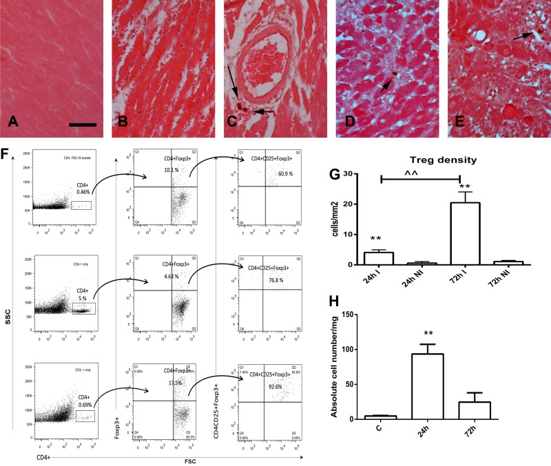
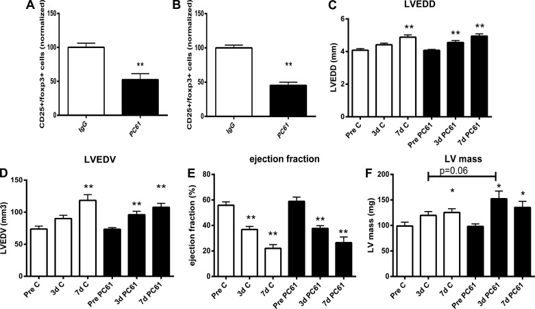
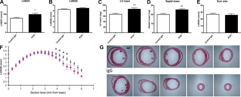
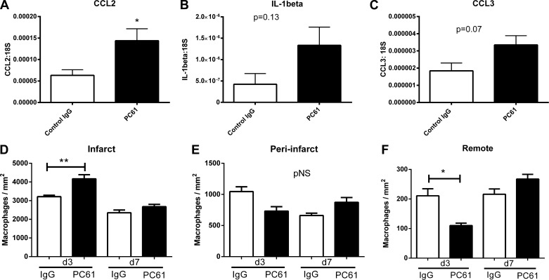
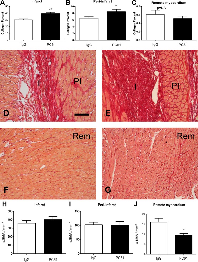
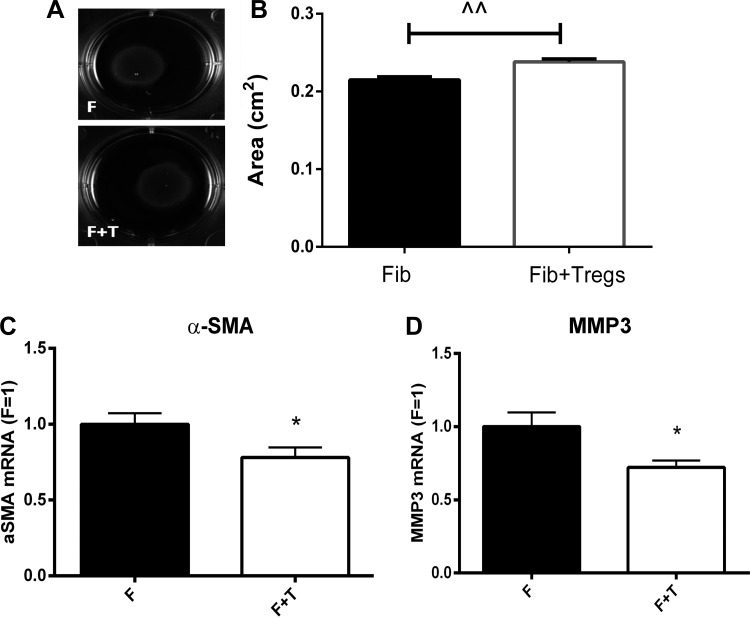
Regulatory T cells (Tregs) play a pivotal role in suppressing immune responses regulating behavior and gene expression in effector T cells, macrophages, and dendritic cells. Tregs infiltrate the infarcted myocardium; however, their role the inflammatory and reparative response after myocardial infarction remains poorly understood. We used FoxP3(EGFP) reporter mice to study Treg trafficking in the infarcted heart and examined the effects of Treg depletion on postinfarction remodeling using an anti-CD25 antibody. Moreover, we investigated the in vitro effects of Tregs on cardiac fibroblast phenotype and function. Low numbers of Tregs infiltrated the infarcted myocardium after 24-72 h of reperfusion. Treg depletion had no significant effects on cardiac dysfunction and scar size after reperfused myocardial infarction but accelerated ventricular dilation and accentuated apical remodeling. Enhanced myocardial dilation in Treg-depleted animals was associated with increased expression of chemokine (C-C motif) ligand 2 and accentuated macrophage infiltration. In vitro, Tregs modulated the cardiac fibroblast phenotype, reducing expression of α-smooth muscle actin, decreasing expression of matrix metalloproteinase-3, and attenuating contraction of fibroblast-populated collagen pads. Our findings suggest that endogenous Tregs have modest effects on the inflammatory and reparative response after myocardial infarction. However, the anti-inflammatory and matrix-preserving properties of Tregs may suggest a role for Treg-based cell therapy in the attenuation of adverse postinfarction remodeling.

Keywords: fibroblast; inflammation; lymphocyte; myocardial infarction; remodeling.


Figures

Fig. 1.
Fig. 1.
Recruitment of regulatory T cells (Tregs) in the infarcted myocardium. Foxp3EGFP mice were used to identify Tregs in the infarcted myocardium using two different strategies: immunohistochemistry (A–E) and flow cytometry (F). Immunohistochemical staining for EGFP showed no Tregs in noninfarcted mouse hearts (A); occasional perivascular and interstitial Tregs were identified (arrows) in infarcted hearts after 24 h (B and C) and 72 h (D and E) of reperfusion. Scale bar = 15 μm. F: representative flow cytometric analysis demonstrating infiltration of the infarcted myocardium with CD4+CD25+forkhead box P3 (FoxP3)+ Tregs. Cells harvested from control hearts (bottom) and from infarcted hearts after 24 h (middle) or 72 h (top) of reperfusion were fluorescently labeled for CD4 and then gated for FoxP3. Finally, the number of CD4+CD25+FoxP3+ Tregs was quantitated. G: quantitative analysis of histological sections showing significantly higher numbers of Tregs in infarcted segments (I) versus noninfarcted segments (NI) after 24 and 72 h of reperfusion. **P < 0.01 vs. corresponding NI; ^^P < 0.01 vs. 24 h. H: quantitation of flow cytometric analysis showing the marked increase in the number of CD4+CD25+FoxP3+ Tregs in the infarct after 24 h of reperfusion. **P < 0.01 vs. control (C) hearts.
Fig. 2.
Fig. 2.
Systemic administration of anti-CD25 antibody (PC61) was used to deplete Tregs in C57BL6J mice. The effectiveness of the strategy was tested using flow cytometry to quantitatively assess Treg numbers in the mouse spleen. PC61 administration resulted in a marked reduction in splenic Tregs 3 days (A) and 5 days (B) after injection. **P < 0.01 vs. IgG control (n = 7–8/group). Echocardiographic assessment showed that in both PC61- and IgG-treated animals, infarcted hearts showed chamber dilation [as evidenced by increased left ventricular (LV) end-diastolic diameter (LVEDD; C) and LV end-diastolic volume (LVEDV; D)], systolic dysfunction (as suggested by reduced ejection fraction; E), and increased LV mass (F) after 3–7 days of reperfusion. *P < 0.05 and **P < 0.01 vs. corresponding baseline values. Treg depletion had no significant effect on ventricular dilation and systolic dysfunction after myocardial infarction.
Fig. 3.
Fig. 3.
Quantitative morphometric analysis of perfusion-fixed hearts using systematic reconstruction of the ventricular geometry from the base to apex was used to study the effects of Treg depletion on adverse postinfarction remodeling 7 days after coronary occlusion-reperfusion. Treg depletion significantly increased morphometrically derived LVEDV (A; *P < 0.05 vs. the IgG-treated group) but did not significantly affect LVEDD (B; defined as the maximal internal dimension of the ventricle). LV mass (C) and septal mass (D) were significantly increased in Treg-depleted animals (**P < 0.01); however, scar size was comparable between groups (E). F: assessment of regional ventricular remodeling by measuring LV end-diastolic internal dimension in sections obtained at 300-μm partitions from the base to apex showing that Treg depletion accentuated apical remodeling without affecting the maximal dimension of the ventricle (blue, PC61; and red, control IgG). G: representative images of infarcted hearts from a PC61-treated animal (top) and an IgG-treated animal (bottom) at five different levels are shown to illustrate the apical dilation observed in the absence of Tregs. Scale bar = 1 mm.
Fig. 4.
Fig. 4.
Treg depletion accentuates the postinfarction inflammatory response. Compared with mice that received control IgG, animals treated with PC61 antibody exhibited increased monocyte chemotactic protein-1/chemokine (C-C motif) ligand (CCL)2 mRNA levels (A; *P < 0.05) and had a trend toward increased expression of IL-1β (B) and CCL3 (C). D: macrophage density in the infarcted region was higher in PC61-treated mice after 3 days of reperfusion. In contrast, no significant differences were noted in macrophage density in the peri-infarct area (E), whereas macrophage density was lower in the remote noninfarcted myocardium (F) of Treg-depleted animals. *P < 0.05 and **P < 0.01 vs. the corresponding IgG-treated group. pNS, P = not significant.
Fig. 5.
Fig. 5.
Treg depletion is associated with modestly increased collagen content in the infarcted area without affecting myofibroblast density. A–C: quantitative analysis of the collagen-stained area in the infarct (I; A), peri-infarct zone (PI; B), and remote remodeling myocardium (Rem; C) after 7 days of reperfusion. Treg depletion increased collagen content in the infarct and peri-infarct zone but did not affect collagen levels in the remote remodeling myocardium. *P < 0.05 and **P < 0.01 vs. control IgG. D–G: representative photomicrographs of collagen-stained sections from control IgG-treated (D and F) and PC61-treated (E and G) animals showing identification of the collagen network in the infarct, peri-infarct region, and remote remodeling myocardium. H–J: quantitative analysis of myofibroblast density in the infarcted myocardium (H), peri-infarct zone (I), and remote remodeling myocardium (J). Treg depletion did not affect myofibroblast density in the infarcted and peri-infarct areas but was associated with reduced myofibroblast density in the remote remodeling myocardium. *P < 0.05 vs. IgG. Scale bar = 50 μm.
Fig. 6.
Fig. 6.
Tregs reduce contraction of collagen pads by cardiac fibroblasts (Fib). Cardiac fibroblasts were cocultured with Tregs to study the effects of Tregs on the contraction of fibroblast-populated collagen pads. A and B: coculture with Tregs was associated with a modest but highly reproducible reduction in collagen pad contraction (F, fibroblasts; F + T, fibroblasts + Tregs). ^^P < 0.01. C and D: attenuated collagen pad contraction was associated with reduced α-SMA mRNA expression (C) and decreased matrix metalloproteinase (MMP)-3 synthesis (D). *P < 0.05 (n = 10–14).

References

    1. Bujak M, Dobaczewski M, Gonzalez-Quesada C, Xia Y, Leucker T, Zymek P, Veeranna V, Tager AM, Luster AD, Frangogiannis NG. Induction of the CXC chemokine interferon-γ-inducible protein 10 regulates the reparative response following myocardial infarction. Circ Res 105: 973–983, 2009 - PMC - PubMed
    1. Bujak M, Ren G, Kweon HJ, Dobaczewski M, Reddy A, Taffet G, Wang XF, Frangogiannis NG. Essential role of Smad3 in infarct healing and in the pathogenesis of cardiac remodeling. Circulation 116: 2127–2138, 2007 - PubMed
    1. Chandrasekar B, Smith JB, Freeman GL. Ischemia-reperfusion of rat myocardium activates nuclear factor-κB and induces neutrophil infiltration via lipopolysaccharide-induced CXC chemokine. Circulation 103: 2296–2302, 2001 - PubMed
    1. Chaudhry A, Rudensky AY. Control of inflammation by integration of environmental cues by regulatory T cells. J Clin Invest 123: 939–944, 2013 - PMC - PubMed
    1. Chen W, Saxena A, Li N, Sun J, Gupta A, Lee DW, Tian Q, Dobaczewski M, Frangogiannis NG. Endogenous IRAK-M attenuates postinfarction remodeling through effects on macrophages and fibroblasts. Arterioscler Thromb Vasc Biol 32: 2598–2608, 2012 - PMC - PubMed

MeSH terms