. 2015 Mar;21(3):292-302.
doi: 10.1089/ten.TEC.2014.0296. Epub 2014 Sep 19.

Application and assessment of optical clearing methods for imaging of tissue-engineered neural stem cell spheres

Affiliations

Application and assessment of optical clearing methods for imaging of tissue-engineered neural stem cell spheres

Molly E Boutin et al. Tissue Eng Part C Methods. 2015 Mar.

Abstract

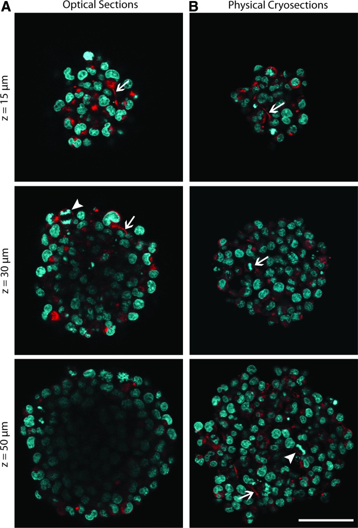
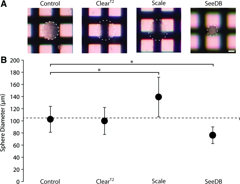
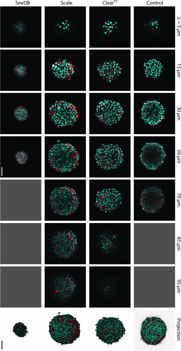
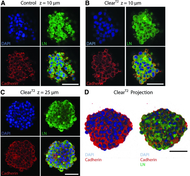
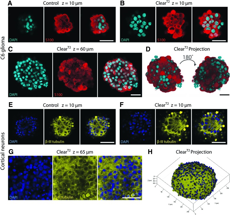
Three-dimensional (3D) cell culture is an important tool that facilitates biological discoveries by bridging the divide between standard two-dimensional cell culture and the complex, high-cell-density in vivo environment. Typically, the internal structures of 3D tissue-engineered samples are visualized through an involved process of physical sectioning, immunostaining, imaging, and computational reconstruction. However, recent progress in tissue-clearing methods has improved optical-imaging-depth capabilities in whole embryos and brains by reducing tissue opacity and light scattering, thus decreasing the need for physical sectioning. In this study, we assessed the application of the recently published clearing techniques Clear(T2), Scale, and SeeDB to tissue-engineered neural spheres. We found that scaffold-free self-assembled adult hippocampal neural stem cell spheres of 100-μm diameter could be optically cleared and imaged using either Clear(T2) or Scale, while SeeDB only marginally improved imaging depth. The Clear(T2) protocol maintained sphere size, while Scale led to sample expansion, and SeeDB led to sample shrinkage. Additionally, using Clear(T2) we cleared and successfully imaged spheres of C6 glioma cells and spheres of primary cortical neurons. We conclude that Clear(T2) is the most effective protocol of those tested at clearing neural spheres of various cell types and could be applied to better understand neural cell interactions in 3D tissue-engineered samples.


Figures

<b>FIG. 1.</b>
FIG. 1.
Comparison of optical and physical sections of neural stem cell (NSC) spheres shows signal losses in center of optical sections. NSC spheres fixed after 3 days in culture were 104.64±22.01 μm in diameter. (A) Optical sections of immunostained whole spheres in phosphate-buffered saline (PBS), at depths of 15, 30, and 50 μm. (B) Cryosections of 8-μm thickness were immunostained and images show sections of corresponding sizes to optical sections. Immunostaining was performed for nestin (red), and counterstained for DAPI (cyan). Arrows highlight cellular extensions; arrowheads highlight dividing cells. Scale bar indicates 50 μm. Color images available online at www.liebertpub.com/tec
<b>FIG. 2.</b>
FIG. 2.
Changes in transparency and size were observed in 3-day-old NSC spheres after clearing with different methods. (A) Light transmission images of control and cleared spheres imaged on crosshatched backgrounds to show relative differences in transparency. White dotted lines outline spheres. Scale bar indicates 50 μm. (B) Graph of NSC sphere diameters after fixation, immunostaining, and incubation in either control PBS or clearing solutions. Horizontal dotted line indicates average sphere diameter prior to fixation. *p<0.05. Color images available online at www.liebertpub.com/tec
<b>FIG. 3.</b>
FIG. 3.
Confocal slices at denoted z-depths and corresponding projections of 3-day-old NSC spheres show changes in fluorescent nestin (red) and DAPI (cyan) signal visualization due to the application of different clearing protocols. Gray boxes represent depths at which an image could not be acquired. Scale bar indicates 50 μm. Color images available online at www.liebertpub.com/tec
<b>FIG. 4.</b>
FIG. 4.
Extracellular laminin and transmembrane cadherin were preserved after the ClearT2 protocol. NSC spheres were fixed after 3 days in culture, immunostained, DAPI counterstained (blue), and cleared. Comparing superficial optical sections of control (A) and ClearT2-treated (B) spheres demonstrates that laminin (green) and cadherin (red) antigens were preserved after clearing (z=10 μm). (C) Laminin and cadherin staining was visible within the ClearT2-treated sphere (z=25 μm). (D) Z-stack projection of an entire sphere (diameter=76 μm) shows cadherin located at cell–cell junctions and laminin present extracellularly. All scale bars indicate 50 μm. Color images available online at www.liebertpub.com/tec
<b>FIG. 5.</b>
FIG. 5.
ClearT2 protocol was successfully applied to spheres composed of various neural cell types. (A–D) C6 glioma spheres were fixed after 1 day in culture, immunostained, DAPI counterstained (cyan), and cleared. Comparing superficial optical sections of control (A) and ClearT2-treated (B) spheres illustrates that S100 (red) antigen was preserved after clearing (z=10 μm). (C) ClearT2-treated sphere shows S100 staining halfway through the sphere (z=60 μm). (D) 180° rotation of 110-μm z-stack projection demonstrates effective clearing throughout the entire C6 sphere. (E–H) Primary cortical neuron spheres were fixed after 7 days in culture, immunostained, DAPI counterstained (blue), and cleared. Comparing superficial optical sections of control (E) and ClearT2-treated (F) spheres confirms that β-III tubulin (yellow) antigen was preserved after clearing (z=10 μm). (G) ClearT2-treated sphere shows β-III tubulin staining halfway through the sphere (z=65 μm). (H) Z-stack projection of half of a cortical sphere (depth=67 μm) displays β-III tubulin network formation in sphere periphery. All scale bars indicate 50 μm. Color images available online at www.liebertpub.com/tec

References

    1. Nakatomi H., Kuriu T., Okabe S., Yamamoto S., Hatano O., Kawahara N., Tamura A., Kirino T., and Nakafuku M.Regeneration of hippocampal pyramidal neurons after ischemic brain injury by recruitment of endogenous neural progenitors. Cell 110,429, 2002 - PubMed
    1. Salman H., Ghosh P., and Kernie S.G.Subventricular zone neural stem cells remodel the brain following traumatic injury in adult mice. J Neurotrauma 21,283, 2004 - PubMed
    1. Doetsch F.A niche for adult neural stem cells. Curr Opin Genet Dev 13,543, 2003 - PubMed
    1. Temple S.The development of neural stem cells. Nature 414,112, 2001 - PubMed
    1. Ming G.-L., and Song H.Adult neurogenesis in the mammalian brain: significant answers and significant questions. Neuron 70,687, 2011 - PMC - PubMed

Publication types