

PKG-1α leucine zipper domain defect increases pulmonary vascular tone: implications in hypoxic pulmonary hypertension
- PMID: 25128522
- PMCID: PMC4187036
- DOI: 10.1152/ajplung.00093.2014
PKG-1α leucine zipper domain defect increases pulmonary vascular tone: implications in hypoxic pulmonary hypertension
Abstract
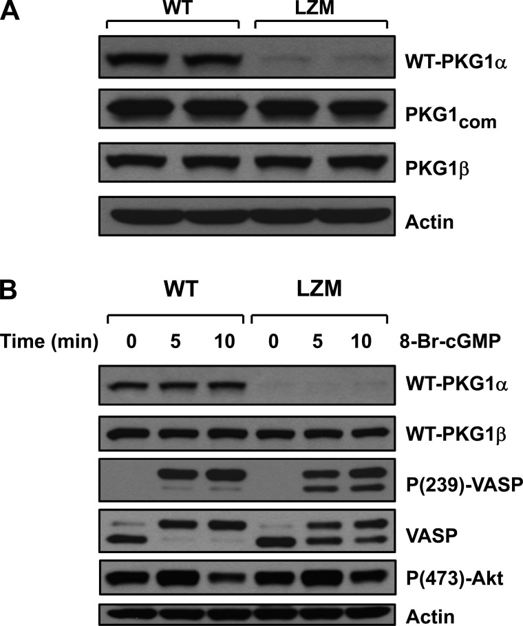
Pulmonary hypertension (PH) is a chronic disease characterized by a progressive increase in vasomotor tone, narrowing of the vasculature with structural remodeling, and increase in pulmonary vascular resistance. Current treatment strategies include nitric oxide therapy and methods to increase cGMP-mediated vasodilatation. cGMP-dependent protein kinases (PKG) are known mediators of nitric oxide- and cGMP-induced vasodilatation. Deletion of PKG-1 in mice has been shown to induce PH, however, the exact mechanisms by which loss of PKG-1 function leads to PH is not known. In a mouse model with a selective mutation in the NH2-terminus leucine zipper protein interaction domain of PKG-1α [leucine zipper mutant (LZM)], we found a progressive increase in right ventricular systolic pressure and right heart hypertrophy compared with wild-type (WT) mice and increased RhoA-GTPase activity in the lungs. When exposed to chronic hypoxia, LZM mice developed modestly enhanced right ventricular remodeling compared with WT mice. Tadalafil, a phosphodiesterase-5 inhibitor that increases cGMP levels, significantly attenuated hypoxia-induced cardiopulmonary remodeling in WT mice but had no effect in LZM mice. We conclude that a functional leucine zipper domain in PKG-1α is essential for maintenance of a low pulmonary vascular tone in normoxia and for cGMP-mediated beneficial effects of phosphodiesterase-5 inhibition in hypoxic cardiopulmonary remodeling.
Keywords: hypoxia; leucine zipper mutant guanosine 3′,5′-cyclic monophosphate-dependent protein kinase; phosphodiesterases; pulmonary hypertension and right ventricular hypertrophy.
Copyright © 2014 the American Physiological Society.
Figures


Similar articles
-
Sustained therapeutic hypercapnia attenuates pulmonary arterial Rho-kinase activity and ameliorates chronic hypoxic pulmonary hypertension in juvenile rats.Am J Physiol Heart Circ Physiol. 2012 Jun 15;302(12):H2599-611. doi: 10.1152/ajpheart.01180.2011. Epub 2012 Apr 13. Am J Physiol Heart Circ Physiol. 2012. PMID: 22505643
-
Attenuated vasodilatation in lambs with endogenous and exogenous activation of cGMP signaling: role of protein kinase G nitration.J Cell Physiol. 2011 Dec;226(12):3104-13. doi: 10.1002/jcp.22692. J Cell Physiol. 2011. PMID: 21351102 Free PMC article.
-
P21-dependent protective effects of a carbon monoxide-releasing molecule-3 in pulmonary hypertension.Arterioscler Thromb Vasc Biol. 2014 Feb;34(2):304-12. doi: 10.1161/ATVBAHA.113.302302. Epub 2013 Dec 12. Arterioscler Thromb Vasc Biol. 2014. PMID: 24334871
-
Mechanisms of pulmonary vascular dysfunction in pulmonary hypertension and implications for novel therapies.Am J Physiol Heart Circ Physiol. 2022 May 1;322(5):H702-H724. doi: 10.1152/ajpheart.00021.2022. Epub 2022 Feb 25. Am J Physiol Heart Circ Physiol. 2022. PMID: 35213243 Free PMC article. Review.
-
Transcriptional and post-transcriptional regulation of cGMP-dependent protein kinase (PKG-I): pathophysiological significance.Cardiovasc Res. 2013 Feb 1;97(2):200-7. doi: 10.1093/cvr/cvs327. Epub 2012 Nov 8. Cardiovasc Res. 2013. PMID: 23139241 Free PMC article. Review.
Cited by
-
Redox regulation of cGMP-dependent protein kinase Iα in the cardiovascular system.Front Pharmacol. 2015 Jul 17;6:139. doi: 10.3389/fphar.2015.00139. eCollection 2015. Front Pharmacol. 2015. PMID: 26236235 Free PMC article. Review.
-
IRAG1 Deficient Mice Develop PKG1β Dependent Pulmonary Hypertension.Cells. 2020 Oct 13;9(10):2280. doi: 10.3390/cells9102280. Cells. 2020. PMID: 33066124 Free PMC article.
-
Smooth Muscle Insulin-Like Growth Factor-1 Mediates Hypoxia-Induced Pulmonary Hypertension in Neonatal Mice.Am J Respir Cell Mol Biol. 2016 Dec;55(6):779-791. doi: 10.1165/rcmb.2015-0388OC. Am J Respir Cell Mol Biol. 2016. PMID: 27438786 Free PMC article.
-
Tetramethylpyrazine Improves Monocrotaline-Induced Pulmonary Hypertension through the ROS/iNOS/PKG-1 Axis.J Healthc Eng. 2022 Mar 24;2022:1890892. doi: 10.1155/2022/1890892. eCollection 2022. J Healthc Eng. 2022. PMID: 35368928 Free PMC article.
-
Update on novel targets and potential treatment avenues in pulmonary hypertension.Am J Physiol Lung Cell Mol Physiol. 2016 Nov 1;311(5):L811-L831. doi: 10.1152/ajplung.00302.2016. Epub 2016 Sep 2. Am J Physiol Lung Cell Mol Physiol. 2016. PMID: 27591245 Free PMC article. Review.
References
-
- Archer SL, Michelakis ED. Phosphodiesterase type 5 inhibitors for pulmonary arterial hypertension. N Engl J Med 361: 1864–1871, 2009 - PubMed
-
- Blanton RM, Takimoto E, Aronovitz M, Thoonen R, Kass DA, Karas RH, Mendelsohn ME. Mutation of the protein kinase I alpha leucine zipper domain produces hypertension and progressive left ventricular hypertrophy: a novel mouse model of age-dependent hypertensive heart disease. J Gerontol A Biol Sci Med Sci 68: 1351–1355, 2013 - PMC - PubMed
-
- Cavasin MA, Demos-Davies K, Horn TR, Walker LA, Lemon DD, Birdsey N, Weiser-Evans MC, Harral J, Irwin DC, Anwar A, Yeager ME, Li M, Watson PA, Nemenoff RA, Buttrick PM, Stenmark KR, McKinsey TA. Selective class I histone deacetylase inhibition suppresses hypoxia-induced cardiopulmonary remodeling through an antiproliferative mechanism. Circ Res 110: 739–748, 2012 - PMC - PubMed
-
- Dumitrascu R, Weissmann N, Ghofrani HA, Dony E, Beuerlein K, Schmidt H, Stasch JP, Gnoth MJ, Seeger W, Grimminger F, Schermuly RT. Activation of soluble guanylate cyclase reverses experimental pulmonary hypertension and vascular remodeling. Circulation 113: 286–295, 2006 - PubMed
Publication types
MeSH terms
Substances
Grants and funding
LinkOut - more resources
Full Text Sources
Other Literature Sources
Medical
Molecular Biology Databases