. 2014 Oct 15;592(20):4591-605.
doi: 10.1113/jphysiol.2014.278192. Epub 2014 Aug 15.

Plasticity in the brainstem vagal circuits controlling gastric motor function triggered by corticotropin releasing factor

Affiliations

Plasticity in the brainstem vagal circuits controlling gastric motor function triggered by corticotropin releasing factor

Kirsteen N Browning et al. J Physiol. .

Abstract

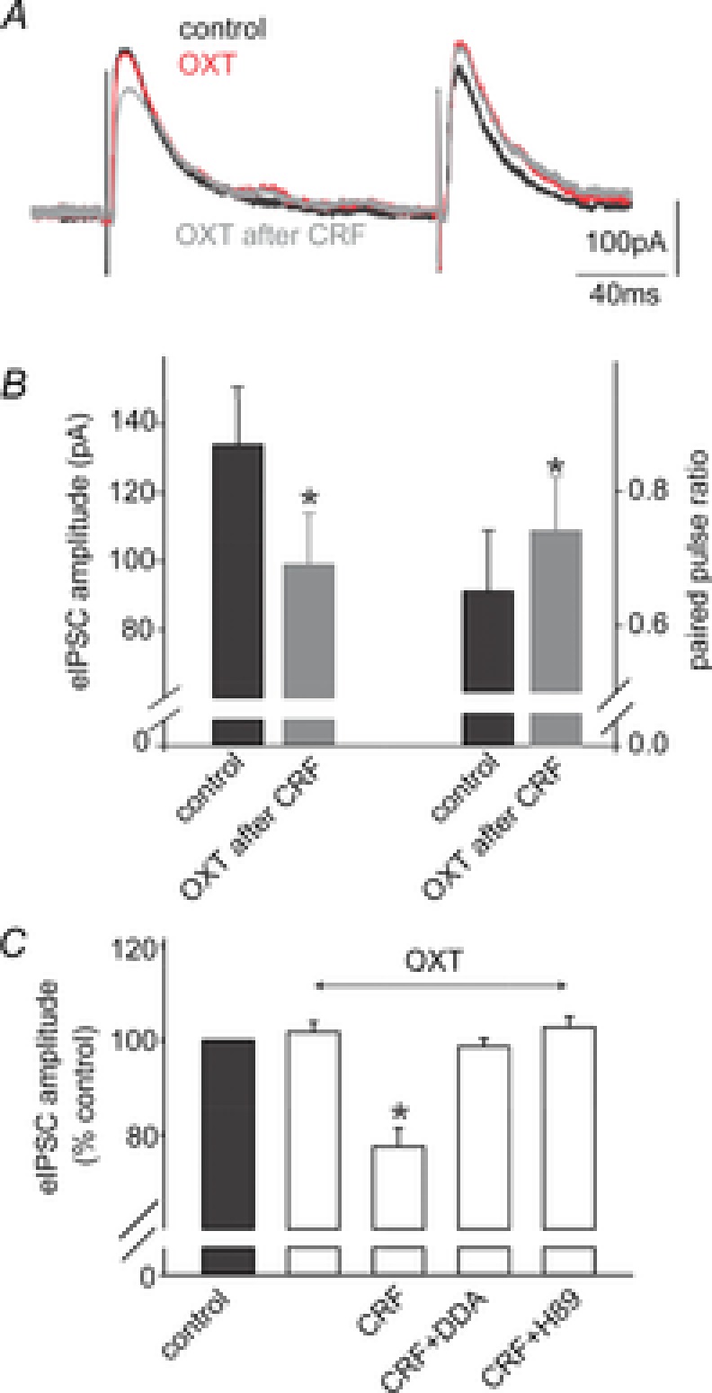
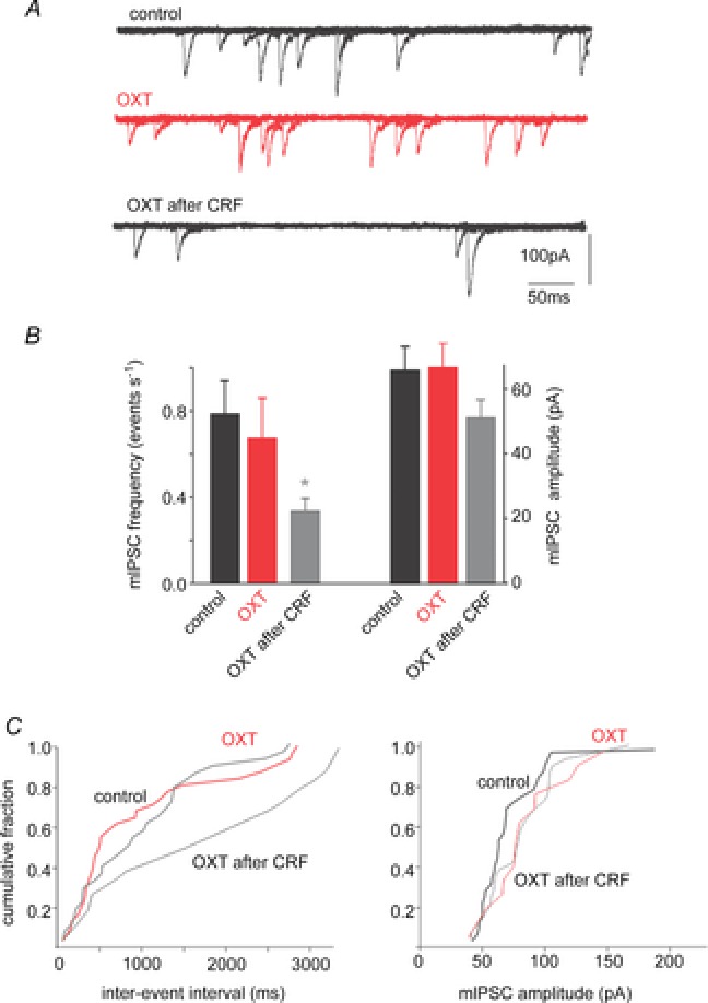
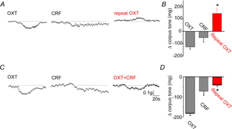
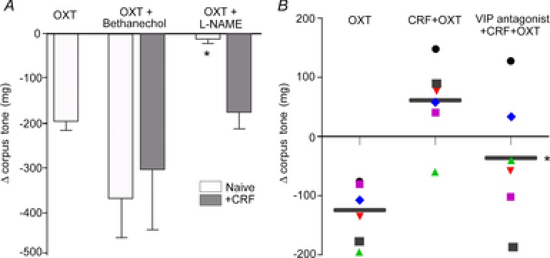
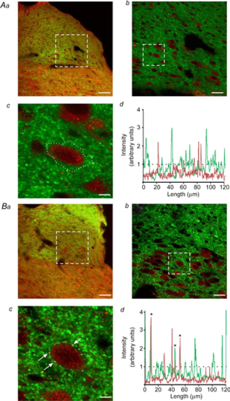
Stress impairs gastric emptying, reduces stomach compliance and induces early satiety via vagal actions. We have shown recently that the ability of the anti-stress neuropeptide oxytocin (OXT) to modulate vagal brainstem circuits undergoes short-term plasticity via alterations in cAMP levels subsequent to vagal afferent fibre-dependent activation of metabotropic glutamate receptors. The aim of the present study was to test the hypothesis that the OXT-induced gastric response undergoes plastic changes in the presence of the prototypical stress hormone, corticotropin releasing factor (CRF). Whole cell patch clamp recordings showed that CRF increased inhibitory GABAergic synaptic transmission to identified corpus-projecting dorsal motor nucleus of the vagus (DMV) neurones. In naive brainstem slices, OXT perfusion had no effect on inhibitory synaptic transmission; following exposure to CRF (and recovery from its actions), however, re-application of OXT inhibited GABAergic transmission in the majority of neurones tested. This uncovering of the OXT response was antagonized by pretreatment with protein kinase A or adenylate cyclase inhibitors, H89 and di-deoxyadenosine, respectively, indicating a cAMP-mediated mechanism. In naive animals, OXT microinjection in the dorsal vagal complex induced a NO-mediated corpus relaxation. Following CRF pretreatment, however, microinjection of OXT attenuated or, at times reversed, the gastric relaxation which was insensitive to l-NAME but was antagonized by pretreatment with a VIP antagonist. Immunohistochemical analyses of vagal motoneurones showed an increased number of oxytocin receptors present on GABAergic terminals of CRF-treated or stressed vs. naive rats. These results indicate that CRF alters vagal inhibitory circuits that uncover the ability of OXT to modulate GABAergic currents and modifies the gastric corpus motility response to OXT.


Figures

Figure 1
Figure 1. Corticotropin-releasing factor uncovers the oxytocin-mediated decrease of the evoked IPSC amplitude via a cAMP/PKA pathway
A, representative traces of evoked IPSCs (eIPSCs) obtained upon electrical stimulation of the adjacent NTS in a gastric-projecting DMV neurone voltage clamped at −50 mV. Perfusion with OXT (100 nm) had no effect upon eIPSCs; however, following perfusion with CRF (100 nm), a second perfusion with OXT reduced the eIPSC amplitude. B, summary graph of the effects of OXT after CRF perfusion on the amplitude of the eIPSC (left) and on the paired pulse ratio (ratio of the amplitude of paired eIPSCs evoked in rapid succession; right). Note that OXT perfusion alters both the eIPSC amplitude as well as the paired pulse ratio. C, summary graph of the normalized effects of OXT on eIPSC amplitude either alone, or after exposure to CRF, CRF + DDA or CRF + H89. Note that the ability of CRF to uncover the effects of OXT were prevented by pretreatment with the adenylate cyclase inhibitor DDA, or the PKA inhibitor H89. *P < 0.05.
Figure 2
Figure 2. Oxytocin-mediated inhibition of miniature inhibitory currents is uncovered by pretreatment with CRF
A, in a gastric-projecting DMV neurone voltage clamped at −50 mV, miniature IPSCs (mIPSCs) were unaffected by perfusion with OXT (100 nm) unless the slice was pretreated with CRF (100 nm). B, summary graph of the effects of OXT, alone and after CRF pretreatment, on the frequency and amplitude of mIPSCs. Note that OXT perfusion reduced the frequency, but did not alter the amplitude significantly, of mIPSCs. *P < 0.05. C, cumulative histograms of the effects of perfusion of OXT, alone and after CRF pretreatment, on the frequency (left) and amplitude (right) of mIPSCs from the neurone illustrated in A.
Figure 3
Figure 3. CRF pretreatment alters the corpus tone response to microinjection of oxytocin in the DVC
Representative trace showing that microinjection of OXT in the DVC decreases corpus tone (left panel). Upon recovery from the OXT-induced corpus relaxation, application of CRF to the floor of the 4th ventricle induced a decrease in gastric tone (middle panel); upon recovery of baseline tone, a second microinjection of OXT increased corpus tone (right panel). The data are summarized in a graphic representation in B. C, representative trace showing that microinjection of OXT in the DVC decreases corpus tone (left panel). Upon recovery from the OXT-induced corpus relaxation, application of CRF to the floor of the 4th ventricle induced a decrease in gastric tone (middle panel); upon recovery of baseline tone, a second microinjection of OXT induced a decrease in corpus tone that was significantly smaller than that obtained upon the first microinjection of OXT (right panel). The data are summarized in a graphic representation in D. *P < 0.05.
Figure 4
Figure 4. Following CRF pretreatment, the oxytocin-induced effects on corpus tone are mediated by VIP
A, bar graphs showing the effects of OXT on the corpus tone in different conditions. Microinjection of OXT in the DVC decreased corpus tone in naive animals (white bar); the effect was prevented by administration of the NO synthase inhibitor l-NAME, but not by the non-selective muscarinic agonist bethanecol. Following pretreatment with CRF, the corpus response to OXT was not inhibited by l-NAME or bethanechol (grey bar). *P < 0.05 vs. OXT alone in naive rats or vs. oxytocin + CRF. B, data points showing the response of individual animals to microinjection of OXT alone (left column), the response to the second administration of OXT conducted after CRF pretreatment (middle column) and the administration of OXT preceded by CRF and VIP antagonist administration (right column). Black bar represents the mean response; P < 0.05 vs. CRF + OXT.
Figure 5
Figure 5. Restraint stress increases the expression of OXT-R on GABAergic terminals apposing DMV neurones
Aa, representative micrograph from a control rat showing GAD-67-IR-positive terminals (green staining) and OXT-R-positive receptors (red staining) in the dorsal vagal complex. Ab, higher magnification of the dotted area in a. Note that DMV neurones are seen as dark areas surrounded by the green immunofluorescence. Ac, single DMV neurone captured at a higher magnification from the area highlighted in b shows no co-localization of GAD-67 puncta (green) with OXT-Rs (red). Ad, graphical representation of colour intensity around the DMV neurone traced in Ac. Metamorph software analysis confirms no overlapping intensity signals between GAD-67 (green line) and OXT-Rs (red line). The dashed line represents the threshold of signal co-localization. Ba, representative micrograph showing GAD-67-IR-positive terminals (green staining) and OXT-R-positive receptors (red staining) in the dorsal vagal complex from a rat that has undergone 2 h of acute restraint stress. Bb, higher magnification of the dotted area in a. Note DMV neurones are seen as dark areas surrounded by green immunofluorescence. Bc, single DMV neurone captured at a higher magnification from the area highlighted in b shows several instances of co-localization of GAD-67 puncta (green) with OXT-Rs (red). Bd, graphical representation of colour intensity around the DMV neurone traced in c. Metamorph software analysis confirms the presence of overlapping intensity signals between GAD-67 (green line) and OXT-Rs (red line). Asterisks in the graph indicate the same co-localized puncta shown in panel c (arrows). The dashed line represents the threshold of signal co-localization. Scale bar: 75 μm for a; 25 μm for b; 5.625 μm for c.

Similar articles

Cited by

References

    1. Babic T, Browning KN, Travagli RA. Differential organization of excitatory and inhibitory synapses within the rat dorsal vagal complex. Am J Physiol Gastrointest Liver Physiol. 2011;300:G21–G32. - PMC - PubMed
    1. Babygirija R, Bulbul M, Cerjak D, Ludwig K, Takahashi T. Sustained acceleration of colonic transit following chronic homotypic stress in oxytocin knockout mice. Neurosci Lett. 2011;495:77–81. - PubMed
    1. Babygirija R, Bulbul M, Yoshimoto S, Ludwig K, Takahashi T. Central and peripheral release of oxytocin following chronic homotypic stress in rats. Auton Neurosci. 2012;167:56–60. - PubMed
    1. Babygirija R, Zheng J, Bulbul M, Cerjak D, Ludwig K, Takahashi T. Sustained delayed gastric emptying during repeated restraint stress in oxytocin knockout mice. J Neuroendocrinol. 2010a;22:1181–1186. - PubMed
    1. Babygirija R, Zheng J, Ludwig K, Takahashi T. Central oxytocin is involved in restoring impaired gastric motility following chronic repeated stress in mice. Am J Physiol Regul Integr Comp Physiol. 2010b;298:R157–R165. - PMC - PubMed

Publication types

MeSH terms

LinkOut - more resources