. 2014 Sep;46(9):1028-33.
doi: 10.1038/ng.3070. Epub 2014 Aug 17.

Jagunal homolog 1 is a critical regulator of neutrophil function in fungal host defense

Affiliations

Jagunal homolog 1 is a critical regulator of neutrophil function in fungal host defense

Gerald Wirnsberger et al. Nat Genet. 2014 Sep.

Abstract

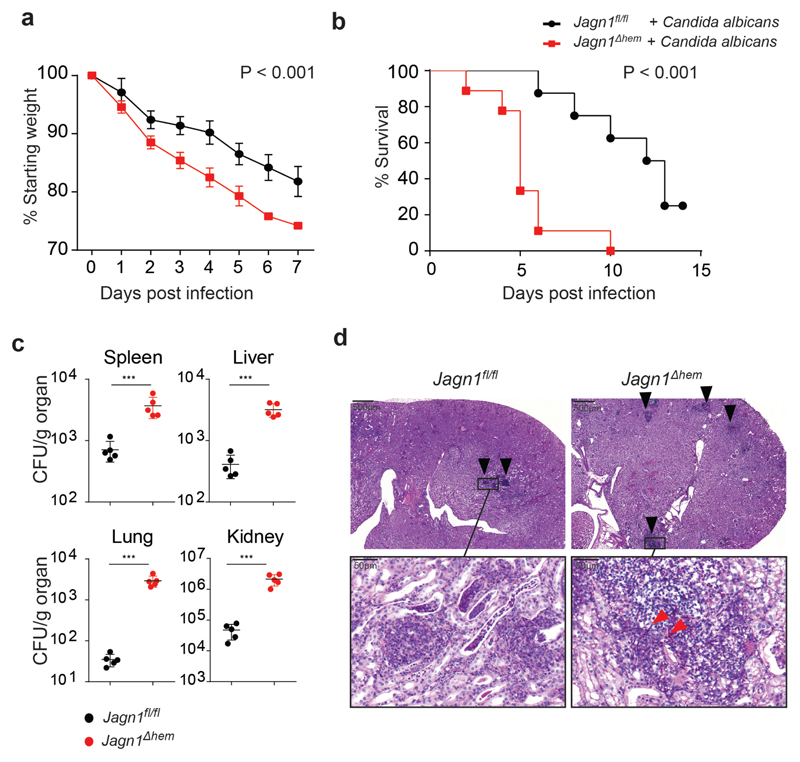
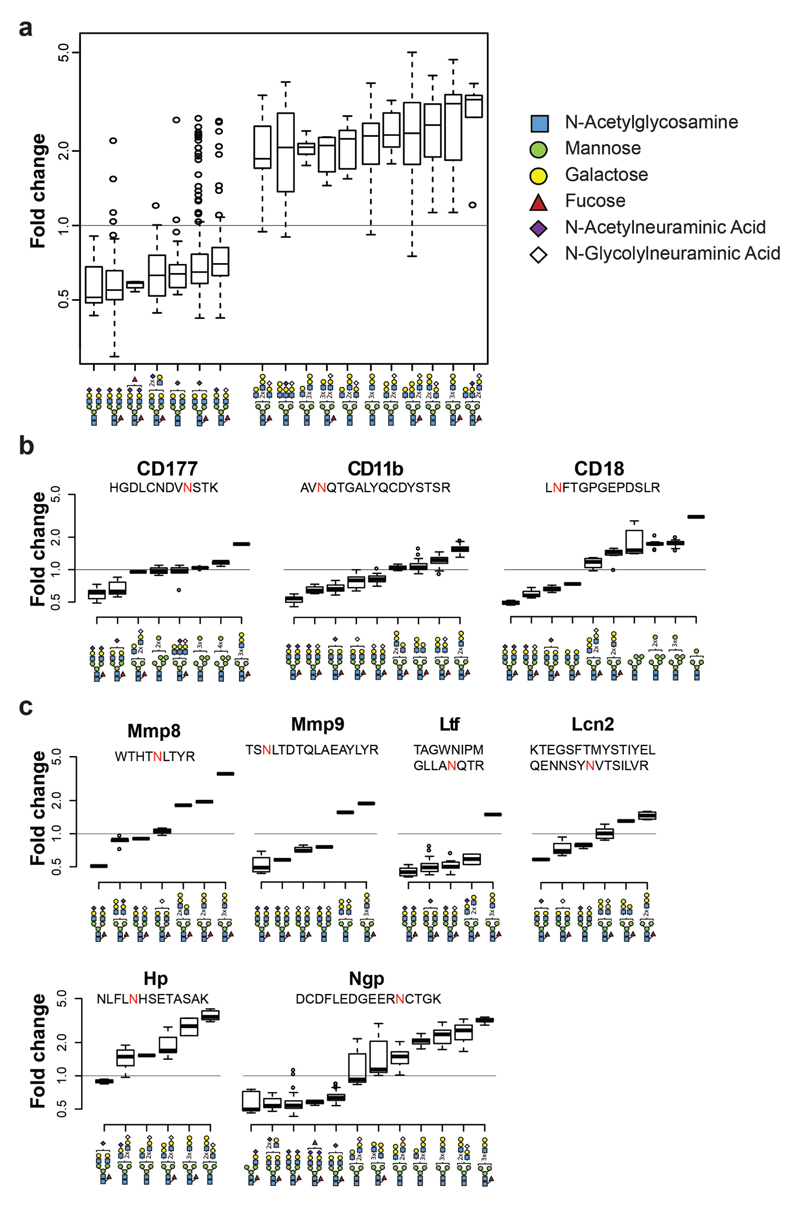
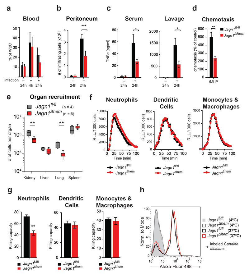
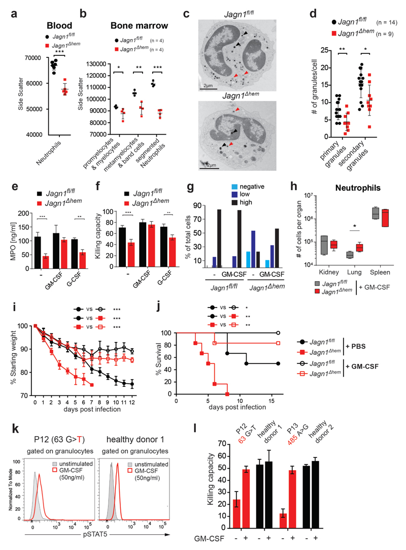
Neutrophils are key innate immune effector cells that are essential to fighting bacterial and fungal pathogens. Here we report that mice carrying a hematopoietic lineage-specific deletion of Jagn1 (encoding Jagunal homolog 1) cannot mount an efficient neutrophil-dependent immune response to the human fungal pathogen Candida albicans. Global glycobiome analysis identified marked alterations in the glycosylation of proteins involved in cell adhesion and cytotoxicity in Jagn1-deficient neutrophils. Functional analysis confirmed marked defects in neutrophil migration in response to Candida albicans infection and impaired formation of cytotoxic granules, as well as defective myeloperoxidase release and killing of Candida albicans. Treatment with granulocyte/macrophage colony-stimulating factor (GM-CSF) protected mutant mice from increased weight loss and accelerated mortality after Candida albicans challenge. Notably, GM-CSF also restored the defective fungicidal activity of bone marrow cells from humans with JAGN1 mutations. These data directly identify Jagn1 (JAGN1 in humans) as a new regulator of neutrophil function in microbial pathogenesis and uncover a potential treatment option for humans.


Conflict of interest statement

Competing Financial Interests

The authors declare no competing financial interests.

Figures

Figure 1
Figure 1. Hematopoietic deletion of Jagn1 impairs neutrophil-dependent immune responses against Candida albicans.
(a,b) Control Jagn1fl/fl and knock-out Jagn1Δhem littermate mice were infected intravenously with Candida albicans (105 CFU/21.5g body weight) and monitored for the indicated time periods. Plots depict (a) weight loss over time after infection as compared to starting weight (p value assessed by two-way ANOVA) or (b) survival over time after infection (p value assessed with log rank test). Data are shown as means ± SD. n = 8 for Jagn1fl/fl and n = 9 for Jagn1Δhem mice. (c) Candida albicans loads in various organs at day 3 after infection are plotted as colony forming units (CFU) per gram organ weight. Data are shown as means ± SD. Each data point represents an individual mouse. *** P <0.001, Student’s t-test. (d) Representative kidney sections from mice 3 days after Candida albicans infection, stained with periodic acid-Schiff. Inflammatory infiltrates are indicated by black arrow heads. Boxed areas in the upper panels (50 x magnification) are shown in the lower panels (600 x magnification). Note the presence of Candida albicans hyphae in the Jagn1Δhem kidney (red arrow heads).
Figure 2
Figure 2. Loss of Jagn1 impairs N-glycosylation of neutrophil homing and effector molecules.
(a) Ratios of N-glycan structures in Jagn1Δhem versus Jagn1fl/fl bone marrow neutrophils that are affected by the loss of Jagn1. The defined glycan structures are indicated. See Supplementary Fig. 4 for complete glyco-proteomic profiles. (b,c) Schematic depiction of N-glycan structures on (b) a selected set of proteins involved in adhesion and migration of neutrophils and (c) proteins involved in tissue remodeling and cytotoxic effector functions of neutrophils that are affected by the loss of Jagn1. Positions of glycosylated asparagines (N) are indicated in red. Shown are the ratios for the indicated glycan structures in Jagn1Δhem versus Jagn1fl/fl neutrophils. Ratios below 1 indicate downregulation in knock-out neutrophils, ratios above 1 show upregulation in Jagn1Δhem knock-out cells. In all panels, data are shown as box-and-whiskers-plots. Horizontal bars indicate the median; boxes span the respective interquantile range; whiskers extend to 1.5 interquantile range; outliers are indicated by circles. n = 3 neutrophils isolated from three different mice for each genotype.
Figure 3
Figure 3. Jagn1 deficient neutrophils exhibit impaired migration and killing of Candida albicans.
(a,b) Jagn1fl/fl and Jagn1Δhem littermate mice were injected intraperitoneally with 5 x 10⁶ CFU of Candida albicans or PBS as a control, and (a) mobilization of neutrophils into the blood stream or (b) recruitment into the peritoneum were measured at the indicated time points. Data are shown as means ± SD. n = 3 per genotype for blood mobilization and n = 6 per genotype for intraperitoneal recruitment. (c) Serum and intraperitoneal lavage levels of TNFα as assayed by ELISA 24 hours after intraperitoneal infection with Candida albicans. Data are shown as mean values ± SD. n = 3 per genotype. (d) fMLP induced chemotaxis of neutrophils. Plot depicts the fMLP induced chemotactic responses of bone marrow neutrophils in a transwell assay. Assays were done in triplicates. Data are shown as mean percentages of chemotaxis as compared to the baseline (set to 100%) ± SD. (e) Control Jagn1fl/fl and knock-out Jagn1Δhem littermate mice were infected intravenously with Candida albicans (105 CFU/21.5g body weight) or PBS as a control and neutrophil organ recruitment was assessed 24 hours later. n = 4 for Jagn1fl/fl and n = 6 for Jagn1Δhem. Whiskers extend to minimum and maximum values, respectively. (f) Reactive oxygen species (ROS) production by bone marrow neutrophils, bone marrow derived dendritic cells, and bone marrow derived monocytes/macrophages co-cultured with Candida albicans and monitored in real time over the indicated time period using the luminol assay. Experiments were performed in triplicates. Values are expressed as relative light units per 1000 cells. (g) Killing capacity of Jagn1fl/fl and Jagn1Δhem bone marrow neutrophils, bone marrow derived dendritic cells, and bone marrow derived monocytes/macrophages as assessed by 24 hour co-culture with Candida albicans. Assays were performed in sextuplicates. (h) Phagocytosis of Candida albicans. Neutrophils of the indicated genotypes were isolated from the bone marrow and co-cultured with Alexa Fluor 488 labeled Candida albicans at either 4°C (negative control) or at 37°C. After 45 minutes of co-culture, neutrophils were analyzed for Candida albicans uptake by flow cytometry. Data are shown as means ± SD. * P <0.05, ** P < 0.01, *** P <0.001 as calculated with Student’s t-test.
Figure 4
Figure 4. Jagn1 controls neutrophil granules and anti-fungal cytotoxicity.
(a,b) Decreased granularity of Jagn1-deficient peripheral blood neutrophils (a) and the indicated bone marrow neutrophil precursor populations (b) as detected by flow cytometry. Each data point represents an individual mouse. (c) Representative electron micrographs of segmented neutrophils isolated from the bone marrow of Jagn1fl/fl and Jagn1Δhem mice. 36000x magnifications. Black arrowheads indicate primary, red arrowheads secondary granules. (d) Quantitation of primary and secondary granule numbers of segmented bone marrow Jagn1fl/fl and Jagn1Δhem neutrophils. (e) MPO release from purified bone marrow neutrophils co-cultured with Candida albicans for 24 hours as measured by ELISA in triplicates with or without recombinant murine GM-CSF or recombinant murine G-CSF (100ng/ml each). (f) Killing capacity of Jagn1fl/fl and Jagn1Δhem bone marrow neutrophils as assessed by 24 hour co-culture of Candida albicans with or without recombinant murine GM-CSF or recombinant murine G-CSF (100ng/ml each). Assays were performed in sextuplicates. (g) MPO expression in neutrophils after in vivo treatment with GM-CSF. Mice of the indicated genotypes were treated daily with recombinant murine GM-CSF (0.5μg) for 3 days or left untreated. MPO expression in blood neutrophils was assessed subsequently by staining with an MPO specific antibody. Neutrophils were scored based on staining intensity as cells expressing no MPO (negative), low levels of MPO (low), and high levels of MPO (high). Plot depicts the distribution of MPO expression levels in neutrophil populations of the indicated genotype with or without GM-CSF treatment (n = 18/32 for Jagn1fl/fl untreated/treated and n = 43/37 for Jagn1Δhem untreated/treated). (h) 24 hours after in vivo GM-CSF pre-treatment control Jagn1fl/fl and knock-out Jagn1Δhem littermate mice were infected intravenously with Candida albicans (105 CFU/21.5g body weight) or PBS and neutrophil recruitment into the indicated organs was assessed 24 hours later. n = 4 for Jagn1fl/fl and n = 5 for Jagn1Δhem mice. Whiskers of Box plots extend to minimum and maximum values, respectively. (i) Weight loss over time after infection as compared to starting weight and (j) survival after infection of Jagn1fl/fl and Jagn1Δhem mice infected i.v. with Candida albicans (105 CFU/21.5g body weight). Mice were treated with either PBS or recombinant murine GM-CSF (0.5μg) every 48 hours (starting 1 day before Candida albicans infection) and monitored for the indicated time periods. n = 6 mice for each cohort. (k) CD34- bone marrow cells from the indicated JAGN1 mutant patient and a healthy donor were stimulated with recombinant human GM-CSF for 15 min and then assayed for phosphorylation of STAT5 (pSTAT5) by intracellular staining. Histogram overlays are shown for cells stimulated with human GM-CSF or left unstimulated. (l) Killing capacity of CD34- bone marrow cells from the indicated JAGN1 mutant patients or healthy controls as assessed by 24 hour co-culture with Candida albicans with or without recombinant human GM-CSF (50ng/ml). Assays were performed in quadruplicates. In all panels except h, data are shown as mean ± SD. P values were calculated using the Student’s t-test with the exception of panel (i) (two-way ANOVA over the entire time period) and panel (j) (log rank test). * P <0.05, ** P < 0.01, *** P <0.001.

Similar articles

Cited by

References

    1. Borregaard N. Neutrophils, from marrow to microbes. Immunity. 2010;33:657–70. - PubMed
    1. Mocsai A. Diverse novel functions of neutrophils in immunity, inflammation, and beyond. J Exp Med. 2013;210:1283–99. - PMC - PubMed
    1. Klein C. Genetic defects in severe congenital neutropenia: emerging insights into life and death of human neutrophil granulocytes. Annu Rev Immunol. 29:399–413. - PubMed
    1. de Boer J, et al. Transgenic mice with hematopoietic and lymphoid specific expression of Cre. Eur J Immunol. 2003;33:314–25. - PubMed
    1. Fulurija A, Ashman RB, Papadimitriou JM. Neutrophil depletion increases susceptibility to systemic and vaginal candidiasis in mice, and reveals differences between brain and kidney in mechanisms of host resistance. Microbiology. 1996;142(Pt 12):3487–96. - PubMed

Publication types

Substances