

doi: 10.1007/s10858-014-9854-y.
Epub 2014 Aug 17.
Effective strategy to assign ¹H- ¹⁵N heteronuclear correlation NMR signals from lysine side-chain NH3₃⁺ groups of proteins at low temperature
Affiliations
- PMID: 25129623
- PMCID: PMC4160661
- DOI: 10.1007/s10858-014-9854-y
Item in Clipboard
Effective strategy to assign ¹H- ¹⁵N heteronuclear correlation NMR signals from lysine side-chain NH3₃⁺ groups of proteins at low temperature
J Biomol NMR.
2014 Sep.
Abstract
Recent studies have shown that lysine side-chain NH3(+) groups are excellent probes for NMR investigations of dynamics involving hydrogen bonds and ion pairs relevant to protein function. However, due to rapid hydrogen exchange, observation of (1)H-(15)N NMR cross peaks from lysine NH3(+) groups often requires use of a relatively low temperature, which renders difficulty in resonance assignment. Here we present an effective strategy to assign (1)H and (15)N resonances of NH3(+) groups at low temperatures. This strategy involves two new (1)H/(13)C/(15)N triple-resonance experiments for lysine side chains. Application to a protein-DNA complex is demonstrated.
Figures
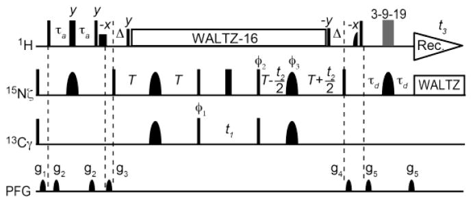
Pulse sequences for the 3D H3NCG experiment for Lys side-chain NH3+ resonance assignment. Thin and bold bars in black represent hard rectangular 90° and 180° pulses, respectively. Unless indicated otherwise, pulse phases are along x. Carrier positions: 1H, the position of the water resonance; 15N, 33 ppm; and 13C, 20 ppm. Short-bold bars represent water-selective soft-rectangular 1H 90° pulses (1.2 ms). The 3-9-19 pulse train-based WATERGATE scheme (Piotto et al. 1992) was used to suppress the water signal. The 13Cγ-selective pulses should not affect 13Cε nuclei (~42ppm). RF strengths for 1H and 15N WALTZ-16 composite pulses (Shaka et al. 1983) were 3.3 kHz and 1.0 kHz, respectively. Shaped pulses: 1H half-Gaussian 90° pulse (2.1 ms); 13C I-BURP2 180° pulse (1.2 ms); and 15N r-SNOB 180° pulse (1.03 ms). Delays: τa = 2.7 ms; δ = 2.6 ms; and T = 50 ms. Phase cycles: ϕ1 = [2x, 2(−x)], ϕ2 = [x, −x], ϕ3 = [4x, 4y], and receiver = [x, −x,−x, x, −x, x, x, −x]. Quadrature detections for indirect 13C and 15N dimensions were achieved using States-TPPI (Marion et al. 1989) for ϕ1 and ϕ2, respectively.

Strips of the 3D H3NCG and H3CECD spectra for all Lys side-chain NH3+ groups of the Egr-1 – DNA complex at 10 °C. Negative contours are shown in green. The 1H and 15N spectral widths for were 18.0 ppm and 4.7 ppm, respectively, in both experiments. The 13C spectral widths were 20.7 ppm in H3NCG and 26.0 ppm in H3NCECD. In each experiment, 32 scans were accumulated per FID, and numbers of complex points for 1H, 13C, and 15N dimensions were 810, 32, and 32, respectively. The total experimental time was 60 hours each. These spectra were recorded at the 1H frequency of 750 MHz using a Bruker Avance III spectrometer equipped with a non-cryogenic TXI probe.

The pulse sequence of the 2D (H2C)N(CC)H-TOCSY experiment. This experiment provides correlation between Lys side-chain 15Nζ and 1H resonances, and corresponds to a hybrid of the H2CN (Andre et al. 2007) and HCCH-TOCSY (Kay et al. 1993) experiments for H2O samples. 13C shaped pulses are Lys 13Cε-selective r-SNOB pulses (980 us). Length of the 13C- spin lock (SL) was 1 ms. 13C carrier position was 43 ppm. The rf strength of the DIPSI-3 scheme (the total length, 16.9 ms) was 9.6 kHz and that of hard 13C pulses was 20.8 kHz. 15N shaped pulses are Lys 15Nζ-selective r-SNOB pulses (1.0 ms). 15N carrier position was 32 ppm. Delays: τ1 = 1.6 ms; τ2 = 1.1 ms; δ = 1.7 ms; and Tc = 20 ms. Phase cycles: ϕ1 = [x, −x]; ϕ2 = [2x, 2(−x)], ϕ3 = [4x, 4y]; ϕ4 = [4x, 4(−x)]; and receiver = [x, −x, −x, x, −y, y, y, −y]. Quadrature detections for indirect 15N dimensions was achieved using States-TPPI for ϕ1.

Use of 2D (H2C)N(CCH)-TOCSY spectrum and temperature dependence of 2D H2(C)N spectra for assignment of Lys NH3+ groups. The spectra were recorded for 0.8 mM 13C/15N-labeled Egr-1 – DNA complex at pH 5.8. (A) The lysine NH3+-selective HISQC (Iwahara et al. 2007) spectrum recorded at 10 °C. (B) Lysine side-chain-specific H2(C)N (Andre et al. 2007) spectra recorded at 10 °C (black), 18 °C (blue), 27 °C (magenta), and 35 °C (red). (C) (H2C)N(CC)H-TOCSY spectrum recorded at 35 °C. The spectra widths and numbers of complex points: 13.4 ppm and 400 points for 1H; and 2.2 ppm and 50 points for 15N. The mixing time of the 13C DIPSI-3 scheme (Shaka et al. 1988) was 16.9 ms. 1024 scans were accumulated per FID. The total titme to record the 2D (H2C)N(CC)H-TOCSY spectrum was 56 hours. All spectra shown in this figure were recorded with a Bruker Avance III spectrometer equipped with a QCI cryogenic probe operated at the 1H frequency of 600 MHz.
Similar articles
-
A Unique and Simple Approach to Improve Sensitivity in 15N-NMR Relaxation Measurements for NH₃⁺ Groups: Application to a Protein-DNA Complex.Molecules. 2017 Aug 15;22(8):1355. doi: 10.3390/molecules22081355. Molecules. 2017. PMID: 28809801 Free PMC article.
-
Heteronuclear NMR spectroscopy for lysine NH(3) groups in proteins: unique effect of water exchange on (15)N transverse relaxation.J Am Chem Soc. 2007 Mar 14;129(10):2971-80. doi: 10.1021/ja0683436. Epub 2007 Feb 15. J Am Chem Soc. 2007. PMID: 17300195
-
Dynamics of lysine side-chain amino groups in a protein studied by heteronuclear 1H−15N NMR spectroscopy.J Am Chem Soc. 2011 Feb 2;133(4):909-19. doi: 10.1021/ja107847d. J Am Chem Soc. 2011. PMID: 21186799
-
NMR studies on the dynamics of hydrogen bonds and ion pairs involving lysine side chains of proteins.Adv Protein Chem Struct Biol. 2013;93:37-80. doi: 10.1016/B978-0-12-416596-0.00002-6. Adv Protein Chem Struct Biol. 2013. PMID: 24018322 Review.
-
NMR Methods for Characterizing the Basic Side Chains of Proteins: Electrostatic Interactions, Hydrogen Bonds, and Conformational Dynamics.Methods Enzymol. 2019;615:285-332. doi: 10.1016/bs.mie.2018.08.017. Epub 2018 Sep 27. Methods Enzymol. 2019. PMID: 30638532 Free PMC article. Review.
Cited by
-
Balancing between affinity and speed in target DNA search by zinc-finger proteins via modulation of dynamic conformational ensemble.Proc Natl Acad Sci U S A. 2015 Sep 15;112(37):E5142-9. doi: 10.1073/pnas.1507726112. Epub 2015 Aug 31. Proc Natl Acad Sci U S A. 2015. PMID: 26324943 Free PMC article.
-
A Unique and Simple Approach to Improve Sensitivity in 15N-NMR Relaxation Measurements for NH₃⁺ Groups: Application to a Protein-DNA Complex.Molecules. 2017 Aug 15;22(8):1355. doi: 10.3390/molecules22081355. Molecules. 2017. PMID: 28809801 Free PMC article.
-
Direct detection of lysine side chain NH3+ in protein-heparin complexes using NMR spectroscopy.Analyst. 2018 Feb 7;143(3):635-638. doi: 10.1039/c7an01406f. Epub 2018 Jan 2. Analyst. 2018. PMID: 29292440 Free PMC article.
-
Entropic Enhancement of Protein-DNA Affinity by Oxygen-to-Sulfur Substitution in DNA Phosphate.Biophys J. 2015 Sep 1;109(5):1026-37. doi: 10.1016/j.bpj.2015.07.032. Biophys J. 2015. PMID: 26331260 Free PMC article.
-
A chemical approach for site-specific identification of NMR signals from protein side-chain NH₃⁺ groups forming intermolecular ion pairs in protein-nucleic acid complexes.J Biomol NMR. 2015 May;62(1):1-5. doi: 10.1007/s10858-015-9909-8. Epub 2015 Feb 19. J Biomol NMR. 2015. PMID: 25690740 Free PMC article.
References
-
- Andre I, Linse S, Mulder FA. Residue-specific pKa determination of lysine and arginine side chains by indirect 15N and 13C NMR spectroscopy: application to apo calmodulin. J Am Chem Soc. 2007;129:15805–15813. - PubMed
-
- Blaum BS, Deakin JA, Johansson CM, Herbert AP, Barlow PN, Lyon M, Uhrín D. Lysine and arginine side chains in glycosaminoglycan-protein complexes investigated by NMR, cross-linking, and mass spectrometry: a case study of the factor H-heparin interaction. J Am Chem Soc. 2010;132:6374–81. - PubMed
Publication types
MeSH terms
Substances
Grants and funding
LinkOut - more resources
Full Text Sources
Other Literature Sources
