

Recurrence of solitary fibrous tumor of the cervical spinal cord
- PMID: 25130009
- PMCID: PMC4345722
Recurrence of solitary fibrous tumor of the cervical spinal cord
Abstract
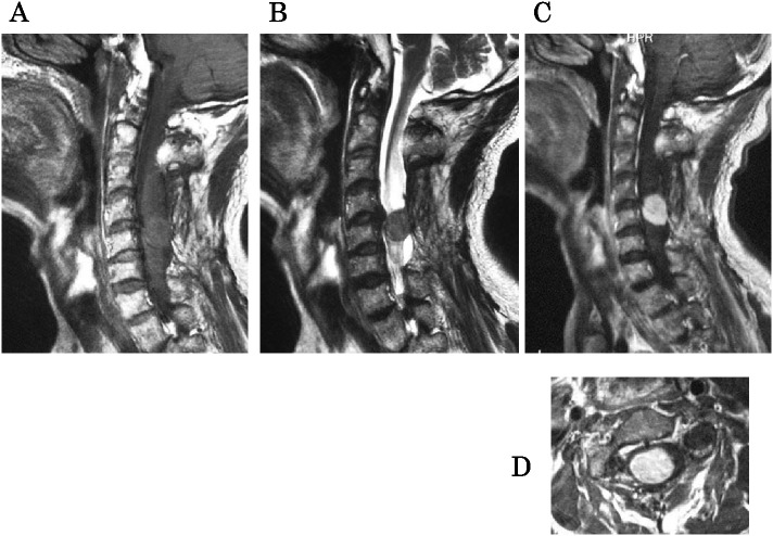
Solitary fibrous tumor (SFT) mostly originates from the pleura because of proliferation of fibroblast cells. It is extremely rare for the tumor to originate from the spinal cord. Here, we report a rare case of SFT in the spinal cord that recurred repeatedly and progressed from intramedullary to extramedullary. A 40-year-old man underwent C4-5 intramedullary and extramedullary tumor resection in another hospital. Eighteen years later, he experienced symptoms of myelopathy because of tumor recurrence; therefore, he consulted with our hospital and underwent tumor resection again. During surgery, we found that the tumor had an intramedullary and extramedullary location. Only partial resection was possible because of intraoperative deterioration in the compound motor action potential (CMAP). After resection, the pathological diagnosis was SFT. The postoperative course was good. However, two years later, a third tumor resection was required because of dysuria and tumor growth. In this surgery, total resection of the tumor was possible without intraoperative deterioration of the CMAP. The tumor has not subsequently recurred. However, SFT recurrence is relatively common and careful follow-up is required for early detection of recurrence, even after successful removal of the tumor.
Figures


Similar articles
-
Solitary fibrous tumour with intramedullary component: case report and review of the literature.Neurol Neurochir Pol. 2014;48(2):144-9. doi: 10.1016/j.pjnns.2013.09.006. Epub 2014 Jan 23. Neurol Neurochir Pol. 2014. PMID: 24821642 Review.
-
Recurrent solitary fibrous tumor of the spinal cord: A case report and literature review.Clin Neuropathol. 2020 Mar/Apr;39(2):86-91. doi: 10.5414/NP301192. Clin Neuropathol. 2020. PMID: 31670648 Review.
-
Intramedullary solitary fibrous tumor of the spinal cord.J Orthop Sci. 2009 Jul;14(4):450-4. doi: 10.1007/s00776-009-1339-6. Epub 2009 Aug 7. J Orthop Sci. 2009. PMID: 19662481 No abstract available.
-
A dumbbell-shaped solitary fibrous tumor of the cervical spinal cord.Yonsei Med J. 2008 Feb 29;49(1):167-70. doi: 10.3349/ymj.2008.49.1.167. Yonsei Med J. 2008. PMID: 18306486 Free PMC article.
-
Hemorrhagic intramedullary solitary fibrous tumor of the conus medullaris: case report.J Neurosurg Spine. 2015 Oct;23(4):438-43. doi: 10.3171/2015.1.SPINE13915. Epub 2015 Jun 26. J Neurosurg Spine. 2015. PMID: 26115022
Cited by
-
Cervical, Intradural Extramedullary Solitary Fibrous Tumor of the Spinal Cord: A Case Report and Review of the Literature.Asian J Neurosurg. 2020 Feb 25;15(1):204-209. doi: 10.4103/ajns.AJNS_213_19. eCollection 2020 Jan-Mar. Asian J Neurosurg. 2020. PMID: 32181204 Free PMC article.
-
Intracranial solitary fibrous tumors: A report of two cases and a review of the literature.Oncol Lett. 2016 Feb;11(2):1057-1060. doi: 10.3892/ol.2015.3985. Epub 2015 Dec 2. Oncol Lett. 2016. PMID: 26893690 Free PMC article.
-
Surgical treatment of primary solitary fibrous tumors involving the pelvic ring.PLoS One. 2018 Nov 27;13(11):e0207581. doi: 10.1371/journal.pone.0207581. eCollection 2018. PLoS One. 2018. PMID: 30481195 Free PMC article.
-
Recurrence of Solitary Fibrous Tumor in the Spinal Cord Following Gross Total and Subtotal Resection: A Case Report of Recurrence 19 Years of Post-total Resection and Systematic Literature Review.NMC Case Rep J. 2024 Oct 24;11:297-303. doi: 10.2176/jns-nmc.2024-0145. eCollection 2024. NMC Case Rep J. 2024. PMID: 39554877 Free PMC article.
-
Dumbbell-Shaped Extramedullary Hemangioblastoma of the Thoracic Spine as a Diagnostic Dilemma of Solitary Fibrous Tumor.Int Med Case Rep J. 2021 Feb 12;14:77-81. doi: 10.2147/IMCRJ.S294759. eCollection 2021. Int Med Case Rep J. 2021. PMID: 33603499 Free PMC article.
References
-
- Klemperer P, Coleman BR. Primary neoplasms of the pleura. A report of five cases. Am J Ind Med. 1992; 22: 1–31. - PubMed
-
- Carneiro SS, Scheithauer BW, Nascimento AG, Hirose T, Davis DH. Solitary fibrous tumor of the meninges: a lesion distinct from fibrous meningioma. A clinicopathologic and immunohistochemical study. Am J Clin Pathol. 1996; 106: 217–224. - PubMed
-
- Alston SR, Francel PC, Jane JA Jr. Solitary fibrous tumor of the spinal cord. Am J Surg Pathol. 1997; 21: 477–483. - PubMed
-
- Malek AM, Weller SJ, Price DL Jr, Madsen JR. Solitary fibrous tumor presenting as a symptomatic intraspinal mass: case report. Neurosurgery. 1997; 40: 844–847. - PubMed
-
- Kanahara T, Hirokawa M, Shimizu M, Terayama K, Nakamura E, Hino Y, Mikawa Y, Manabe T. Solitary fibrous tumor of the spinal cord. Report of a case with scrape cytology. Acta Cytol. 1999; 43: 425–428. - PubMed
Publication types
MeSH terms
LinkOut - more resources
Full Text Sources
Miscellaneous