

Lumisterol is metabolized by CYP11A1: discovery of a new pathway
- PMID: 25130438
- PMCID: PMC4252613
- DOI: 10.1016/j.biocel.2014.08.004
Lumisterol is metabolized by CYP11A1: discovery of a new pathway
Abstract
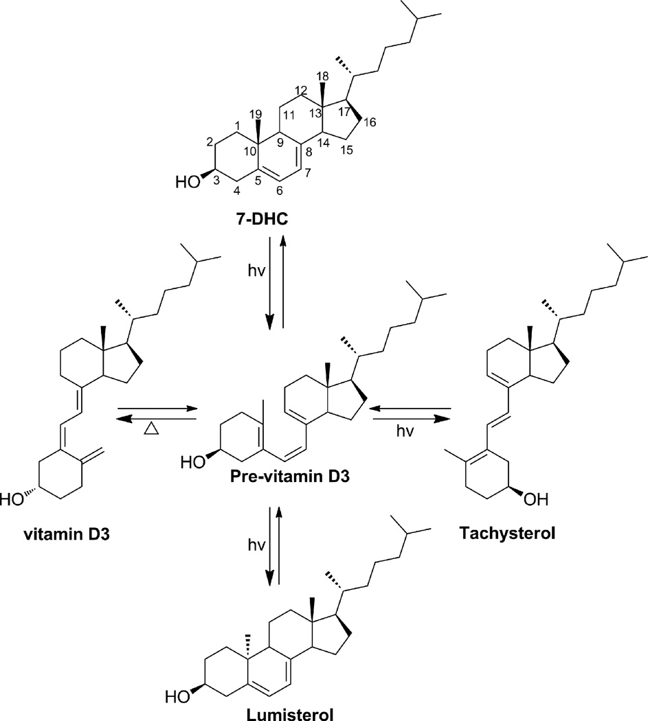
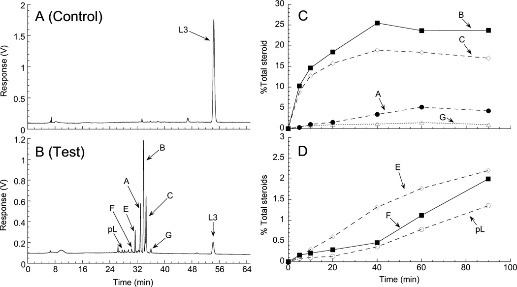
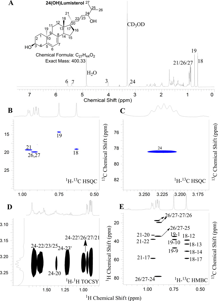
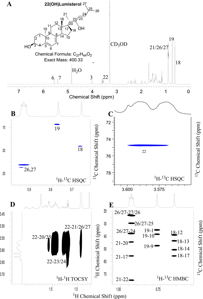
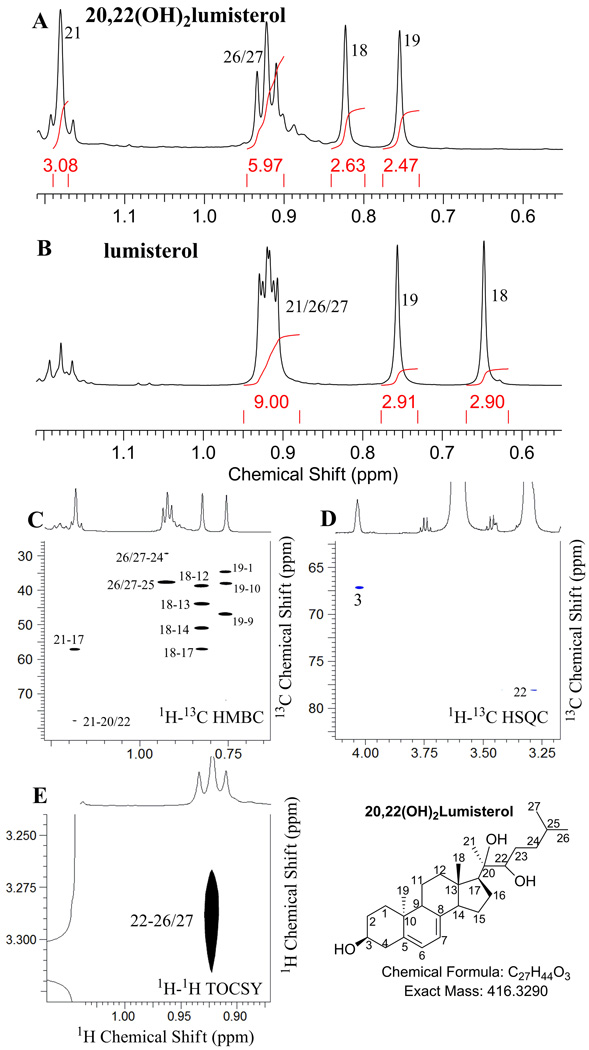
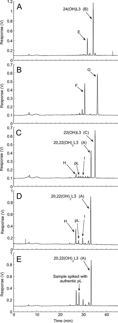
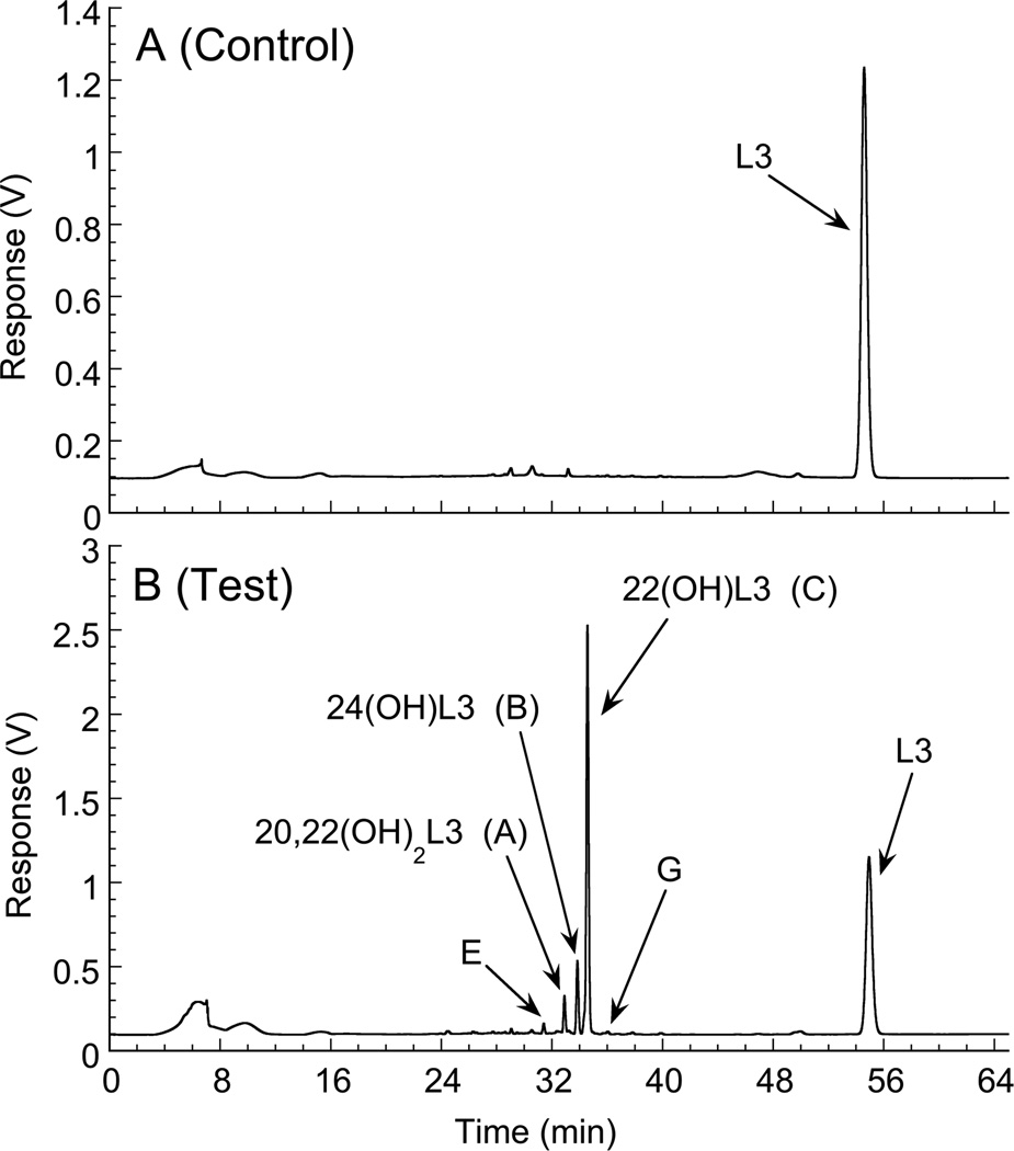
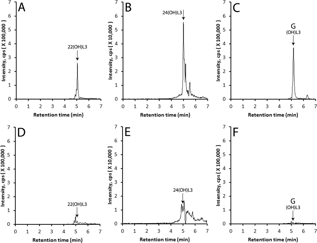
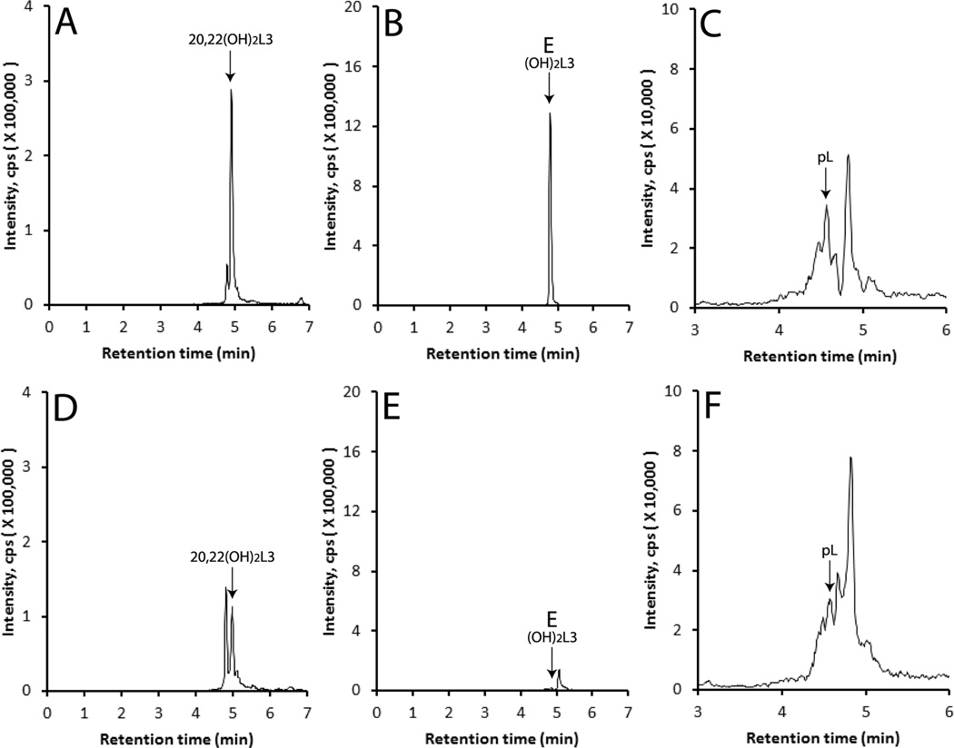
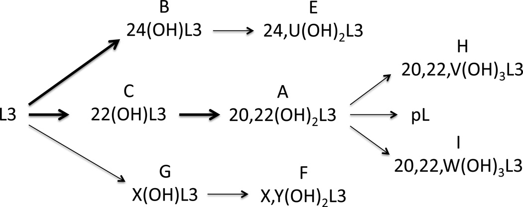
Lumisterol3 (L3) is produced by photochemical transformation of 7-dehydrocholesterol (7-DHC) during exposure to high doses of ultraviolet B radiation. It has been assumed that L3 is biologically inactive and is not metabolized in the body. However, some synthetic derivatives of L3 display biological activity. The aim of this study was to test the ability of CYP11A1 to metabolize L3. Incubation of L3 with bovine or human CYP11A1 resulted in the formation of three major and a number of minor products. The catalytic efficiency of bovine CYP11A1 for metabolism of L3 dissolved in 2-hydroxypropyl-β-cyclodextrin was approximately 20% of that reported for vitamin D3 and cholesterol. The structures of the three major products were identified as 24-hydroxy-L3, 22-hydroxy-L3 and 20,22-dihydroxy-L3 by NMR. 22-Hydroxy-L3 was further metabolized by bovine CYP11A1 to 20,22-dihydroxy-L3. Both 22-hydroxy-L3 and 20,22-dihydroxy-L3 gave rise to a minor metabolite identified from authentic standard and mass spectrometry as pregnalumisterol (pL) (product of C20-C22 side chain cleavage of L3) and two trihydroxy-L3 products. The capability of tissues expressing CYP11A1 to metabolize L3 was demonstrated using pig adrenal fragments where 20,22-dihydroxy-L3, 22-hydroxy-L3, 24-hydroxy-L3 and pL were detected by LC/MS. Thus, we have established that L3 is metabolized by CYP11A1 to 22- and 24-hydroxy-L3 and 20,22-dihydroxy-L3 as major products, as well as to pL and other minor products. The previously reported biological activity of pL and the presence of CYP11A1 in skin suggest that this pathway may serve to produce biologically active products from L3, emphasizing a novel role of CYP11A1 in sterol metabolism.
Keywords: CYP11A1; Cytochrome P450scc; Hydroxylation; Lumisterol; Vitamin D3.
Copyright © 2014 Elsevier Ltd. All rights reserved.
Figures
Similar articles
-
Metabolism of Lumisterol2 by CYP27A1.J Steroid Biochem Mol Biol. 2023 Oct;233:106370. doi: 10.1016/j.jsbmb.2023.106370. Epub 2023 Jul 25. J Steroid Biochem Mol Biol. 2023. PMID: 37499840
-
Human cytochrome P450scc (CYP11A1) catalyzes epoxide formation with ergosterol.Drug Metab Dispos. 2012 Mar;40(3):436-44. doi: 10.1124/dmd.111.042515. Epub 2011 Nov 21. Drug Metab Dispos. 2012. PMID: 22106170 Free PMC article.
-
CYP27A1 acts on the pre-vitamin D3 photoproduct, lumisterol, producing biologically active hydroxy-metabolites.J Steroid Biochem Mol Biol. 2018 Jul;181:1-10. doi: 10.1016/j.jsbmb.2018.02.008. Epub 2018 Feb 13. J Steroid Biochem Mol Biol. 2018. PMID: 29452159 Free PMC article.
-
Photoprotective Properties of Vitamin D and Lumisterol Hydroxyderivatives.Cell Biochem Biophys. 2020 Jun;78(2):165-180. doi: 10.1007/s12013-020-00913-6. Epub 2020 May 22. Cell Biochem Biophys. 2020. PMID: 32441029 Free PMC article. Review.
-
Novel activities of CYP11A1 and their potential physiological significance.J Steroid Biochem Mol Biol. 2015 Jul;151:25-37. doi: 10.1016/j.jsbmb.2014.11.010. Epub 2014 Nov 13. J Steroid Biochem Mol Biol. 2015. PMID: 25448732 Free PMC article. Review.
Cited by
-
Transcriptome Analysis and Identification of the Cholesterol Side Chain Cleavage Enzyme BbgCYP11A1 From Bufo bufo gargarizans.Front Genet. 2022 Apr 5;13:828877. doi: 10.3389/fgene.2022.828877. eCollection 2022. Front Genet. 2022. PMID: 35480310 Free PMC article.
-
Editorial: Steroids and Secosteroids in the Modulation of Inflammation and Immunity.Front Immunol. 2021 Dec 20;12:825577. doi: 10.3389/fimmu.2021.825577. eCollection 2021. Front Immunol. 2021. PMID: 34987528 Free PMC article. No abstract available.
-
The Over-Irradiation Metabolite Derivative, 24-Hydroxylumister-ol3, Reduces UV-Induced Damage in Skin.Metabolites. 2023 Jun 21;13(7):775. doi: 10.3390/metabo13070775. Metabolites. 2023. PMID: 37512482 Free PMC article.
-
Vitamin D and lumisterol derivatives can act on liver X receptors (LXRs).Sci Rep. 2021 Apr 13;11(1):8002. doi: 10.1038/s41598-021-87061-w. Sci Rep. 2021. PMID: 33850196 Free PMC article.
-
Endogenously produced nonclassical vitamin D hydroxy-metabolites act as "biased" agonists on VDR and inverse agonists on RORα and RORγ.J Steroid Biochem Mol Biol. 2017 Oct;173:42-56. doi: 10.1016/j.jsbmb.2016.09.024. Epub 2016 Sep 28. J Steroid Biochem Mol Biol. 2017. PMID: 27693422 Free PMC article. Review.
References
-
- Clarke MW, Tuckey RC, Gorman S, Holt B, Hart PH. Optimized 25-hydroxyvitamin D analysis using liquid-liquid extraction with 2D separation with LC/MS/MS detection, provides superior precision compared to conventional assays. Metabolomics. 2013;9:1031–1040.
-
- De Caprio J, Yun J, Javitt NB. Bile acid and sterol solubilization in 2-hydroxypropyl-β-cyclodextrin. J Lipid Res. 1992;33:441–443. - PubMed
-
- Dixon KM, Norman AW, Sequeira VB, Mohan R, Rybchyn MS, Reeve VE, Halliday GM, Mason RS. 1alpha,25(OH)-vitamin D and a nongenomic vitamin D analogue inhibit ultraviolet radiation-induced skin carcinogenesis. Cancer Prev Res. 2011;4:1485–1494. - PubMed
-
- Holick MF. Vitamin D: a millennium perspective. J Cell Biochem. 2003;88:296–307. - PubMed
Publication types
MeSH terms
Substances
Grants and funding
LinkOut - more resources
Full Text Sources
Other Literature Sources
Molecular Biology Databases
