. 2014 Aug 21;8(4):933-9.
doi: 10.1016/j.celrep.2014.07.003. Epub 2014 Aug 14.

Evidence against a stem cell origin of new hepatocytes in a common mouse model of chronic liver injury

Affiliations

Evidence against a stem cell origin of new hepatocytes in a common mouse model of chronic liver injury

Johanna R Schaub et al. Cell Rep. .

Erratum in

  • Cell Rep. 2014 Sep 11;8(5):1607

Abstract

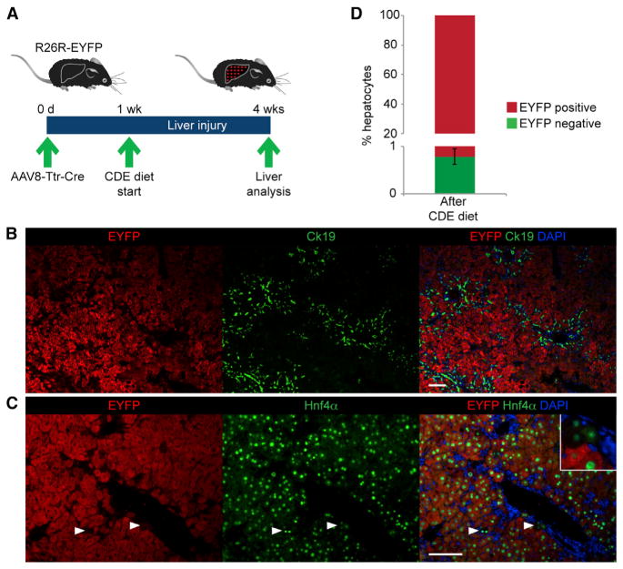
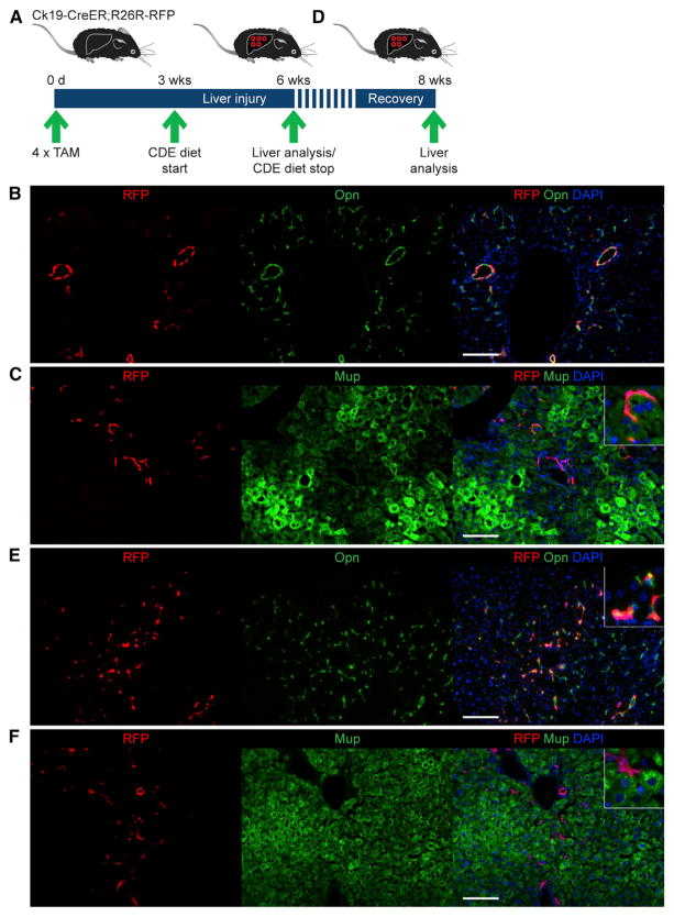
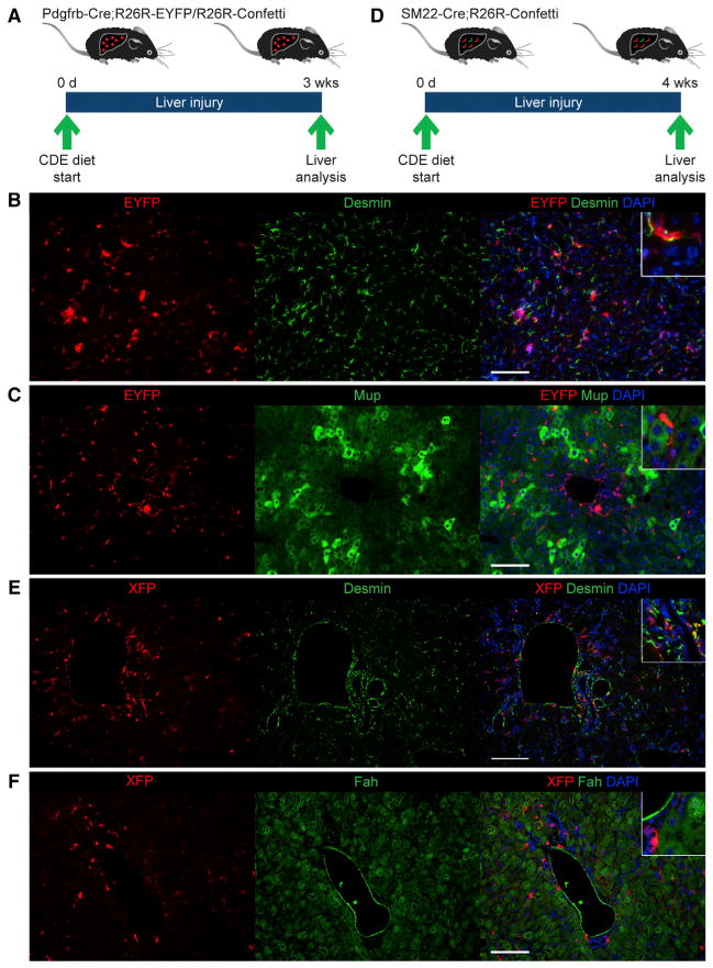
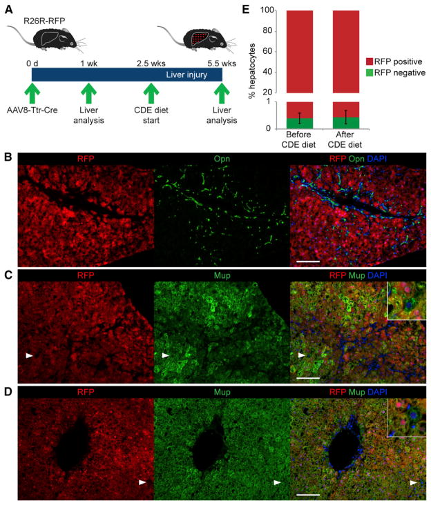
Hepatocytes provide most liver functions, but they can also proliferate and regenerate the liver after injury. However, under some liver injury conditions, particularly chronic liver injury where hepatocyte proliferation is impaired, liver stem cells (LSCs) are thought to replenish lost hepatocytes. Conflicting results have been reported about the identity of LSCs and their contribution to liver regeneration. To address this uncertainty, we followed candidate LSC populations by genetic fate tracing in adult mice with chronic liver injury due to a choline-deficient, ethionine-supplemented diet. In contrast to previous studies, we failed to detect hepatocytes derived from biliary epithelial cells or mesenchymal liver cells beyond a negligible frequency. In fact, we failed to detect hepatocytes that were not derived from pre-existing hepatocytes. In conclusion, our findings argue against LSCs, or other nonhepatocyte cell types, providing a backup system for hepatocyte regeneration in this common mouse model of chronic liver injury.


Figures

Figure 1
Figure 1. Presence of Unlabeled Hepatocytes in a Hepatocyte Fate-Tracing Mouse Model after Chronic Liver Injury
(A) The hepatocyte fate-tracing model was generated by injecting R26R-EYFP mice with AAV8-Ttr-Cre. Liver injury was induced by CDE diet feeding 1 week later. Livers were analyzed after 3 weeks of CDE diet feeding. (B) Coimmunostaining for EYFP and Ck19 shows oval cell expansion characteristic for livers of mice after CDE diet feeding. (C) Coimmunostaining for EYFP and Hnf4α shows EYFP-negative, Hnf4α-positive cells; i.e., non-fate-traced hepatocytes (arrowheads and inset). (D) Quantification of non-fate-traced hepatocytes. Data are shown as mean ± SEM. Scale bars, 100 μm. Representative images and results from three mice are shown. See also Figures S1 and S4B.
Figure 2
Figure 2. Absence of BEC-Derived Hepatocytes
(A) The BEC fate-tracing model was generated by injecting Ck19-CreER;R26R-RFP mice with 4 mg TAM every other day for four injections total. Liver injury was induced by CDE diet feeding 2 weeks after the last TAM injection. Livers were analyzed after 3 weeks of CDE diet feeding. (B) Direct fluorescence of RFP combined with immunostaining for Opn shows no RFP-positive, Opn-negative cells; i.e., non-BECs/nonoval cells derived from BECs. (C) Direct fluorescence of RFP combined with immunostaining for Mup shows no RFP-positive, Mup-positive cells; i.e., hepatocytes derived from BECs. (D) The CDE-Stop model was generated as in (A) with the difference that the mice were fed a normal diet between CDE diet feeding and analysis. (E) Direct fluorescence of RFP combined with immunostaining for Opn shows no RFP-positive, Opn-negative cells; i.e., non-BECs/nonoval cells derived from BECs. (F) Direct fluorescence of RFP combined with immunostaining for Mup shows noRFP-positive, Mup-positive cells; i.e., hepatocytes derived from BECs. Scale bars, 100 μm. At least three mice were analyzed for each experiment. Representative images are shown. See also Figure S2.
Figure 3
Figure 3. Absence of Mesenchymal Liver-Cell-Derived Hepatocytes
(A) Pdgfrb-Cre;R26R-EYFP or Pdgfrb-Cre;R26R-Confetti mice were used for fate tracing of stellate cells/myofibroblasts. Livers were analyzed after 3 weeks of CDE diet feeding. (B) Coimmunostaining for EYFP and desmin shows no EYFP-positive, desmin-negative cells; i.e., nonmesenchymal cells derived from stellate cells/myofibroblasts. (C) Coimmunostaining for EYFP and Mup shows no EYFP-positive, Mup-positive cells; i.e., hepatocytes derived from stellate cells/myofibroblasts. (D) SM22-Cre;R26R-Confetti mice were used for fate tracing of periportal mesenchymal cells. Livers were analyzed after 4 weeks of CDE diet feeding. (E) Coimmunostaining for YFP/nGFP/mCFP (XFP) and desmin shows that desmin-positive periportal mesenchymal cells, but no cells with hepatocyte morphology, are XFP positive. (F) Coimmunostaining for YFP/nGFP/mCFP (XFP) and Fah shows no XFP-positive, Fah-positive cells; i.e., hepatocytes derived from periportal mesenchymal cells. Scale bars, 100 μm. At least three mice were analyzed for each experiment. See also Figure S3.
Figure 4
Figure 4. Origin of New Hepatocytes from Pre-Existing Hepatocytes
(A) A refined hepatocyte fate-tracing model was generated by injecting AAV8-Ttr-Cre into R26R-RFP mice and analyzing the liver of each mouse before and after CDE diet feeding. For this, mice were subjected to one-third PH 1 week after AAV8-Ttr-Cre injection, followed by a 1.5-week-long recovery phase. Final liver analysis was carried out after 3 weeks of CDE diet feeding. (B) Direct fluorescence of RFP combined with immunostaining for Opn shows the characteristic oval cell response after CDE diet feeding. (C and D) Direct fluorescence of RFP combined with immunostaining for Mup shows in livers of mice after (C) and before (D) liver injury the presence of RFP-negative, Mup-positive cells; i.e., non-fate-traced hepatocytes (arrowheads and insets). (E) Quantification of non-fate-traced hepatocytes in livers of mice before and after liver injury. Data are shown as mean ± SEM; p > 0.1. Scale bars, 100 μm. Representative images and results from three mice are shown. See also Figure S4.

References

    1. Akhurst B, Croager EJ, Farley-Roche CA, Ong JK, Dumble ML, Knight B, Yeoh GC. A modified choline-deficient, ethionine-supplemented diet protocol effectively induces oval cells in mouse liver. Hepatology. 2001;34:519–522. - PubMed
    1. Choi TY, Ninov N, Stainier DR, Shin D. Extensive conversion of hepatic biliary epithelial cells to hepatocytes after near total loss of hepatocytes in zebrafish. Gastroenterology. 2014;146:776–788. - PMC - PubMed
    1. Dellavalle A, Sampaolesi M, Tonlorenzi R, Tagliafico E, Sacchetti B, Perani L, Innocenzi A, Galvez BG, Messina G, Morosetti R, et al. Pericytes of human skeletal muscle are myogenic precursors distinct from satellite cells. Nat Cell Biol. 2007;9:255–267. - PubMed
    1. Dorrell C, Erker L, Schug J, Kopp JL, Canaday PS, Fox AJ, Smirnova O, Duncan AW, Finegold MJ, Sander M, et al. Prospective isolation of a bipotential clonogenic liver progenitor cell in adult mice. Genes Dev. 2011;25:1193–1203. - PMC - PubMed
    1. Duncan AW, Dorrell C, Grompe M. Stem cells and liver regeneration. Gastroenterology. 2009;137:466–481. - PMC - PubMed

Publication types