Review
. 2015 May;61(5):1740-6.
doi: 10.1002/hep.27376. Epub 2015 Mar 23.

Liver sinusoidal endothelial cells in hepatic fibrosis

Affiliations
Review

Liver sinusoidal endothelial cells in hepatic fibrosis

Laurie D DeLeve. Hepatology. 2015 May.

Erratum in

  • Correction.
    [No authors listed] [No authors listed] Hepatology. 2015 Jul;62(1):326. doi: 10.1002/hep.27878. Hepatology. 2015. PMID: 26108875 No abstract available.

Abstract

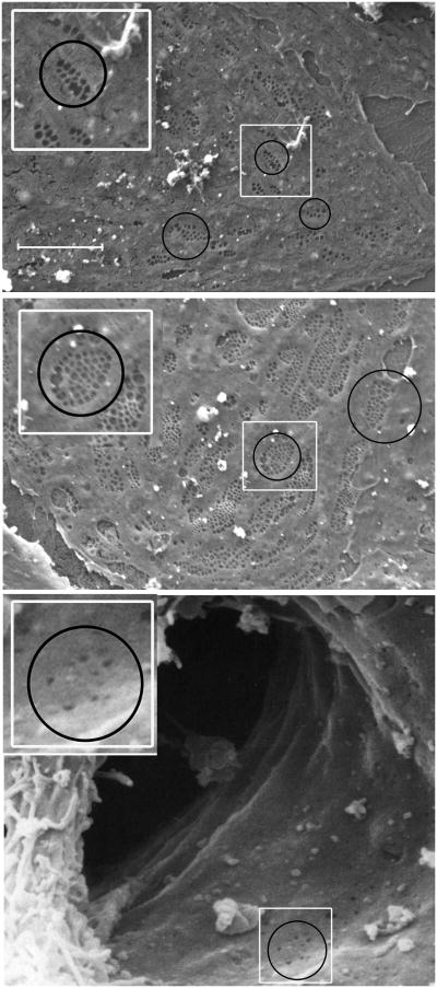
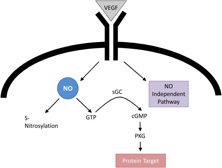
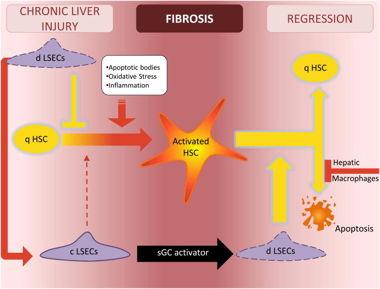
Capillarization, lack of liver sinusoidal endothelial cell (LSEC) fenestration, and formation of an organized basement membrane not only precedes fibrosis, but is also permissive for hepatic stellate cell activation and fibrosis. Thus, dysregulation of the LSEC phenotype is a critical step in the fibrotic process. Both a vascular endothelial growth factor (VEGF)-stimulated, nitric oxide (NO)-independent pathway and a VEGF-stimulated NO-dependent pathway are necessary to maintain the differentiated LSEC phenotype. The NO-dependent pathway is impaired in capillarization and activation of this pathway downstream from NO restores LSEC differentiation in vivo. Restoration of LSEC differentiation in vivo promotes HSC quiescence, enhances regression of fibrosis, and prevents progression of cirrhosis.


Figures

Figure 1
Figure 1. Scanning electron microscopy of liver sinusoidal endothelial cells
Top panel, photomicrograph of a periportal LSEC isolated from rat liver (magnification 5000×). Middle panel, photomicrograph of a centrilobular LSEC isolated form rat liver (magnification 5000×). Note that periportal LSECs (top panel) have fewer fenestrae per sieve plate (average 8 fenestrae per sieve plate), but larger fenestrae than centrilobular LSECs (average 24 fenestrae per sieve plate; middle panel) (2). Smaller, non-clustered holes are endocytic vesicles. Bottom panel, scanning electron microscopic photograph of the lumen of a sinusoid opening into the central vein taken from human liver (magnification 8500×). The sinusoid is bisected by a Kupffer cell. Fenestrae are rare in the endothelial cells that line this transitional zone. Circles surround examples of sieve plates (top two panels) and one of the few areas of the transitional endothelial cell that still has fenestrae (bottom panel). The white rectangles in each panel are enlarged and reproduced in the top left of each panel for better visualization. Bottom panel reproduced from Die Leber des Menschen / The Human Liver Rasterelektronenmikroskopischer Atlas / A Scanning Electron Microscopic Atlas by Franz Vonnahme (ISBN: 978-3-8055-5585-2) with kind permission from S. Karger AG, Basel.
Figure 2
Figure 2. VEGF pathways in LSEC
Maintenance of the LSEC phenotype requires both the VEGF-stimulated NO pathway working through soluble guanylate cyclase, cGMP and protein kinase G and a VEGF pathway working independent of NO. The protein S-nitrosylation pathway is not necessary to maintain the LSEC phenotype. Abbreviations: LSEC, liver sinusoidal endothelial cell; NO, nitric oxide; sGC, soluble guanylate cyclase; VEGF, vascular endothelial growth factor;
Figure 3
Figure 3. The crosstalk of liver cells in regulating HSC quiescence and activation
In chronic liver injury, formation of apoptotic bodies, oxidative stress and cytokines promote HSC activation. Differentiated LSECs function as a gatekeeper, preventing activation of the HSC. However once the LSEC capillarizes, it no longer prevents HSC activation and permits or perhaps promotes activation of the HSC. LSEC capillarization can be reversed with an sGC activator that works downstream in the VEGF-NO-sGC pathway. Reversal of capillarized LSECs to differentiated LSECs promotes reversion of activated HSC to quiescence and induces some apoptosis of activated HSC. Hepatic macrophages in turn inhibit apoptosis of activated HSC. Abbreviations: c LSEC, capillarized liver sinusoidal endothelial cell; d LSEC, differentiated liver sinusoidal endothelial cell; q HSC, quiescent hepatic stellate cell; NO, nitric oxide; sGC, soluble guanylate cyclase.

References

    1. Schaffner F, Popper H. Capillarization of hepatic sinusoids in man. Gastroenterology. 1963;44:239–242. - PubMed
    1. Xie G, L W, W X, W L, DeLeve LD. Isolation of periportal, mid-lobular and centrilobular rat liver sinusoidal endothelial cells enables study of zonated drug toxicity. Am J Physiol-Gastrointest Liver Physiol. 2010;299:G1204–1210. - PMC - PubMed
    1. Sorensen KK, McCourt P, Berg T, Crossley C, Le Couteur D, Wake K, Smedsrod B. The scavenger endothelial cell: a new player in homeostasis and immunity. Am J Physiol Regul Integr Comp Physiol. 2012;303:R1217–1230. - PubMed
    1. Malovic I, Sorensen KK, Elvevold KH, Nedredal GI, Paulsen S, Erofeev AV, Smedsrod BH, et al. The mannose receptor on murine liver sinusoidal endothelial cells is the main denatured collagen clearance receptor. Hepatology. 2007;45:1454–1461. - PubMed
    1. Martens JH, Kzhyshkowska J, Falkowski-Hansen M, Schledzewski K, Gratchev A, Mansmann U, Schmuttermaier C, et al. Differential expression of a gene signature for scavenger/lectin receptors by endothelial cells and macrophages in human lymph node sinuses, the primary sites of regional metastasis. Journal of Pathology. 2006;208:574–589. - PubMed

Publication types