. 2014 Nov 20;515(7527):427-30.
doi: 10.1038/nature13715. Epub 2014 Aug 17.

Structure of malaria invasion protein RH5 with erythrocyte basigin and blocking antibodies

Affiliations

Structure of malaria invasion protein RH5 with erythrocyte basigin and blocking antibodies

Katherine E Wright et al. Nature. .

Abstract

Invasion of host erythrocytes is essential to the life cycle of Plasmodium parasites and development of the pathology of malaria. The stages of erythrocyte invasion, including initial contact, apical reorientation, junction formation, and active invagination, are directed by coordinated release of specialized apical organelles and their parasite protein contents. Among these proteins, and central to invasion by all species, are two parasite protein families, the reticulocyte-binding protein homologue (RH) and erythrocyte-binding like proteins, which mediate host-parasite interactions. RH5 from Plasmodium falciparum (PfRH5) is the only member of either family demonstrated to be necessary for erythrocyte invasion in all tested strains, through its interaction with the erythrocyte surface protein basigin (also known as CD147 and EMMPRIN). Antibodies targeting PfRH5 or basigin efficiently block parasite invasion in vitro, making PfRH5 an excellent vaccine candidate. Here we present crystal structures of PfRH5 in complex with basigin and two distinct inhibitory antibodies. PfRH5 adopts a novel fold in which two three-helical bundles come together in a kite-like architecture, presenting binding sites for basigin and inhibitory antibodies at one tip. This provides the first structural insight into erythrocyte binding by the Plasmodium RH protein family and identifies novel inhibitory epitopes to guide design of a new generation of vaccines against the blood-stage parasite.


Figures
Extended Data Figure 1
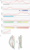
Extended Data Figure 1. PfRH5 disorder predictions and structural alignment
a, Long-range disorder was predicted by POODLE-L and was used to determine domain boundaries for the PfRH5ΔNL crystallisation construct. The disorder predictions are shown above the sequence of PfRH5, with values of >0.5 indicative of disorder. The residues visible in the PfRH5ΔNL crystal structure are shown below the PfRH5 sequence as secondary structure elements (sheets as blue arrows, and helices as tubes in rainbow coloring) linked by blue lines. The missing loop (248-296) is shown as a break in the blue line. The secretion signal sequence is indicated (black underline). b, Two copies of PfRH5 from the PfRH5:basigin structure (red and orange), two copies from the PfRH5:QA1 structure (blue and cyan), and one copy from PfRH5:9AD4 (green) structure were aligned using Coot, giving an RMSD of 1.7 Å. The C-terminus and the loop between helices 4 and 5 were the only regions showing significant differences. For the remaining 95% of PfRH5, the RMSD is 0.9 Å.
Extended Data Figure 2
Extended Data Figure 2. Investigation of the interaction of PfRH5ΔNL with a panel of mouse mAbs using ELISA
Five mAbs that bind to PfRH5 were coated on a ELISA plate and probed using PfRH5ΔNL at concentrations of 12.5, 50, 200 or 800 ng/ml. Antibodies 9AD5, QA1 and QA5 interacted with PfRH5ΔNL while RB3 and 4BA7 did not. Indeed, RB3 and 4BA7 bind to the flexible N-terminus and the truncated loop, respectively, both features lacking in PfRH5ΔNL. The error bars are standard error of mean (n=3).
Extended Data Figure 3
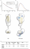
Extended Data Figure 3. SAXS analysis of the PfRH5:basigin complex
a, The theoretical scattering calculated from the average of 20 ab initio reconstructions (continuous lines, with PfRH5 in orange and PfRH5:basigin in blue) plotted with the experimental scattering intensity curves (diamonds). The data are presented as the natural logarithm of the intensity. Guinier plots are displayed in the inset. b, The distance distribution function, P(r), of PfRH5 (orange) and PfRH5:basigin (blue). c, To the left, the crystal structure of PfRH5ΔNL (yellow) was docked into the average ab initio SAXS envelope of full-length PfRH5 (grey). Extra density corresponding to some or all of the truncated regions is visible at the bottom of the kite-like structure, near the C-terminus. To the right, the crystal structure of PfRH5ΔNL:basigin is docked into the average ab initio SAXS envelope of full-length PfRH5:basigin (grey). PfRH5ΔNL is yellow. In dark blue and cyan are basigin molecules from the two PfRH5ΔNL:basigin complexes in the asymmetric unit, superimposed based on the structure of PfRH5ΔNL. d, Summary of SAXS parameters. The radius of gyration (Rg) was determined from the Guinier plot using AutoRg, and the maximum particle dimension (Dmax) and the Porod volume were calculated using GNOM. An estimate of the molecular weight was obtained by dividing the Porod volume by 1.7. Ab initio modeling was used to generate 20 shape reconstructions from the data. The normalised spatial discrepancy parameter (NSD) diagnoses the similarity of these models. The models were averaged and the fit of the average model to the experimental data is indicated by the χ value.
Extended Data Figure 4
Extended Data Figure 4. A conserved PfRH5-like fold in other Plasmodium RH proteins
a, P. falciparum RH1, RH4, RH2b, RH2a and RH3 (a pseudogene); P. vivax RBP-1 and RBP-2; P. reichenowi RH5; and P. yoelii Py01365 were aligned using Clustal Omega and were threaded using the Phyre2 server, giving more than 98% confidence of fold conservation over >260 residues in each case. The secondary structure of PfRH5 is shown below the sequence in a rainbow colour scheme as in Fig. 1A. Residues from PfRH5 that interact with basigin, QA1 and 9AD4 are indicated above the sequence by blue, red or green stars respectively. Cysteine residues that make disulphide bonds are indicated by pink numbers, with residues sharing the same number forming a disulphide bond. b, PfRH5 is shown in yellow, with residues similar among RH proteins (from the alignment in extended data figure 4) highlighted as pink sticks. The majority of the similar residues appear to play a structural role stabilising the architecture of the domain.
Extended Data Figure 5
Extended Data Figure 5. Location of PfRH5 polymorphisms, and residues of PfRH5 and basigin implicated in host tropism
a and b, Indicated are the locations of PfRH5 SNPs that are common (10% frequency or greater; red sticks) or uncommon (blue sticks) among 227 field isolates,,, as well as additional SNPs observed in lab strains (green sticks),. b, Basigin (blue) is shown in addition to PfRH5 (yellow). SNPs Y203, I204, N347, Y358, and E362 are localized in or near the PfRH5:basigin interface. Not visible in this orientation is lab strain polymorphism K429. c, Highlighted are basigin residues F27, Q100, and H102, which affect the affinity for PfRH5 when mutated (orange sticks). Also shown are two SNPs of PfRH5, namely N347 and I204 (pink sticks), found in the PfRH5:basigin binding interface and linked to the strain’s ability to invade Aotus monkey erythrocytes.
Extended Data Figure 6
Extended Data Figure 6. Arrangement of two PfRH5:basigin complexes in the asymmetric unit of the crystal
One complex, shown in yellow (PfRH5) and blue (basigin), interacts with the second, shown in silver (PfRH5) and cyan (basigin), primarily through packing between the two C-terminal domains of basigin. The two C-termini of basigin are in close proximity (top view).
Extended Data Figure 7
Extended Data Figure 7. Analysis of the PfRH5:basigin complex using analytical ultracentrifugation
a-b, Sedimentation velocity analysis. The continuous sedimentation coefficient distributions that best fit the data are shown for basigin (top), full-length PfRH5 (middle), and a gel filtered PfRH5:basigin complex (bottom). The inset shows the fitting residuals. c-d, Sedimentation equilibrium analysis. PfRH5ΔN (residues 140-526), basigin, and a gel filtered PfRH5ΔN:basigin complex were analysed. The runs lasted 20 hours at different speeds, as indicated in the inset legends. UV absorbance was monitored at 280 nm. The residuals are shown below fitted data. The calculated molecular weights are consistent with the formation of a 1:1 complex between PfRH5ΔN and basigin.
Extended Data Figure 8
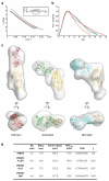
Extended Data Figure 8. SAXS of PfRH5 in complex with growth-inhibitory Fab fragments
a, The theoretical scattering calculated from the average of 20 ab initio reconstructions (continuous lines, with PfRH5 in orange, PfRH5:9AD4 in green, PfRH5:QA1 in red, and PfRH5:QA5 in blue) plotted with the experimental scattering intensity curves (black diamonds). The data are presented as the natural logarithm of the intensity. The Guiner plots are displayed in the inset. b, The distance distribution function, P(r), with colours as in a. c, The crystal structures of PfRH5ΔNL:QA1(left) and PfRH5ΔNL:9AD4 (middle) were docked into the corresponding full-length PfRH5:Fab envelopes (grey). PfRH5ΔNL is shown in yellow, QA1 in red, and 9AD4 in green. PfRH5ΔNL and a Fab fragment (cyan) were docked into the PfRH5:QA5 SAXS envelope to generate a model of the PfRH5:QA5 structure (right). d, A summary of SAXS parameters. The radius of gyration (Rg) was determined from the Guinier plot using AutoRg, and the maximum particle dimension (Dmax) and the Porod volume were calculated using GNOM. An estimate of the molecular weight was obtained by dividing the Porod volume by 1.7. Ab initio modeling was used to generate 20 shape reconstructions from the data. The normalised spatial discrepancy parameter (NSD) diagnoses the similarity of these models. The models were averaged and the fit of the average model to the experimental data is indicated by the χ value.
Figure 1
Figure 1. The structure of PfRH5
a, Three views of PfRH5ΔNL (from the PfRH5ΔNL:9AD4 structure), and a schematic topology diagram, coloured in rainbow from blue (N-terminus) to red (C-terminus). Disulphide bonds are indicated on the topology diagram by red lines. b, PfRH5ΔNL structure docked into a SAXS envelope of full-length PfRH5. c, SPR analysis of the PfRH5ΔNL:basigin interaction. d, In vitro growth inhibition activity (GIA) of IgG from rabbits immunised with PfRH5ΔNL against 3D7 (red) and 7G8 (blue) P. falciparum strains. The error bars are standard error of mean (n=3).
Figure 2
Figure 2. The structure of the PfRH5:basigin complex
a, The structure of PfRH5ΔNL (yellow) bound to basigin (blue). b, A top view of the PfRH5ΔNL:basigin complex showing the two conformations of basigin (blue and cyan) found in the asymmetric unit, aligned on PfRH5ΔNL. c, Equilibrium analytical ultracentrifugation analysis of PfRH5:basigin indicating a 1:1 complex. d, Close-up of the PfRH5:basigin binding site. Basigin residues in the N-terminal domain (pink), the linker (His102, orange stick), and the C-terminal domain (green) contact PfRH5 (grey surface). In the alternative basigin conformation in the asymmetric unit, the cyan loop contacts PfRH5.
Figure 3
Figure 3. Structural analysis of binding of invasion-inhibitory antibody fragments to PfRH5
a, Crystal structures of PfRH5ΔNL (yellow) bound to inhibitory antibody fragments QA1 (red) and 9AD4 (green). Close-up views of the PfRH5 epitopes (red) are shown with antibodies as grey surfaces. b, Top view of PfRH5ΔNL:9AD4 crystal structure with superimposed basigin (blue) aligned on PfRH5. c, Top view of a model of PfRH5:QA5, in a SAXS-derived envelope, with the putative QA5 epitope highlighted red. d, Schematic showing binding sites for the N- and C-terminal domains of basigin (BSG-N and BSG-C; blue), QA1 (red), 9AD4 (green) and QA5 (cyan), on the structure of PfRH5ΔNL.

References

    1. Cowman AF, Crabb BS. Invasion of red blood cells by malaria parasites. Cell. 2006;124:755–766. - PubMed
    1. Tham WH, Healer J, Cowman AF. Erythrocyte and reticulocyte binding-like proteins of Plasmodium falciparum. Trends Parasitol. 2012;28:23–30. - PubMed
    1. Baum J, et al. Reticulocyte-binding protein homologue 5 - an essential adhesin involved in invasion of human erythrocytes by Plasmodium falciparum. Int. J. Parasitol. 2009;39:371–380. - PubMed
    1. Crosnier C, et al. Basigin is a receptor essential for erythrocyte invasion by Plasmodium falciparum. Nature. 2011;480:534–537. - PMC - PubMed
    1. Douglas AD, et al. Neutralization of Plasmodium falciparum merozoites by antibodies against PfRH5. J. Immunol. 2014;192:245–258. - PMC - PubMed

Publication types

MeSH terms