. 2014 Sep 4;513(7516):90-4.
doi: 10.1038/nature13608. Epub 2014 Aug 17.

RIPK1 maintains epithelial homeostasis by inhibiting apoptosis and necroptosis

Affiliations

RIPK1 maintains epithelial homeostasis by inhibiting apoptosis and necroptosis

Marius Dannappel et al. Nature. .

Abstract

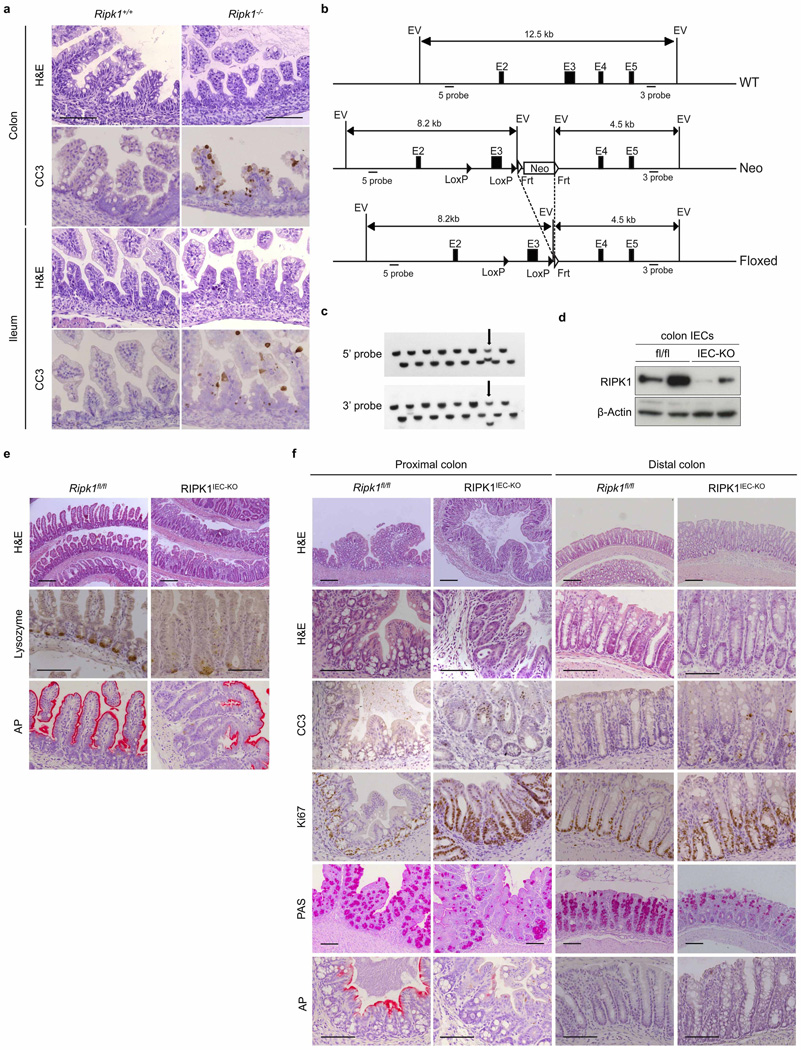
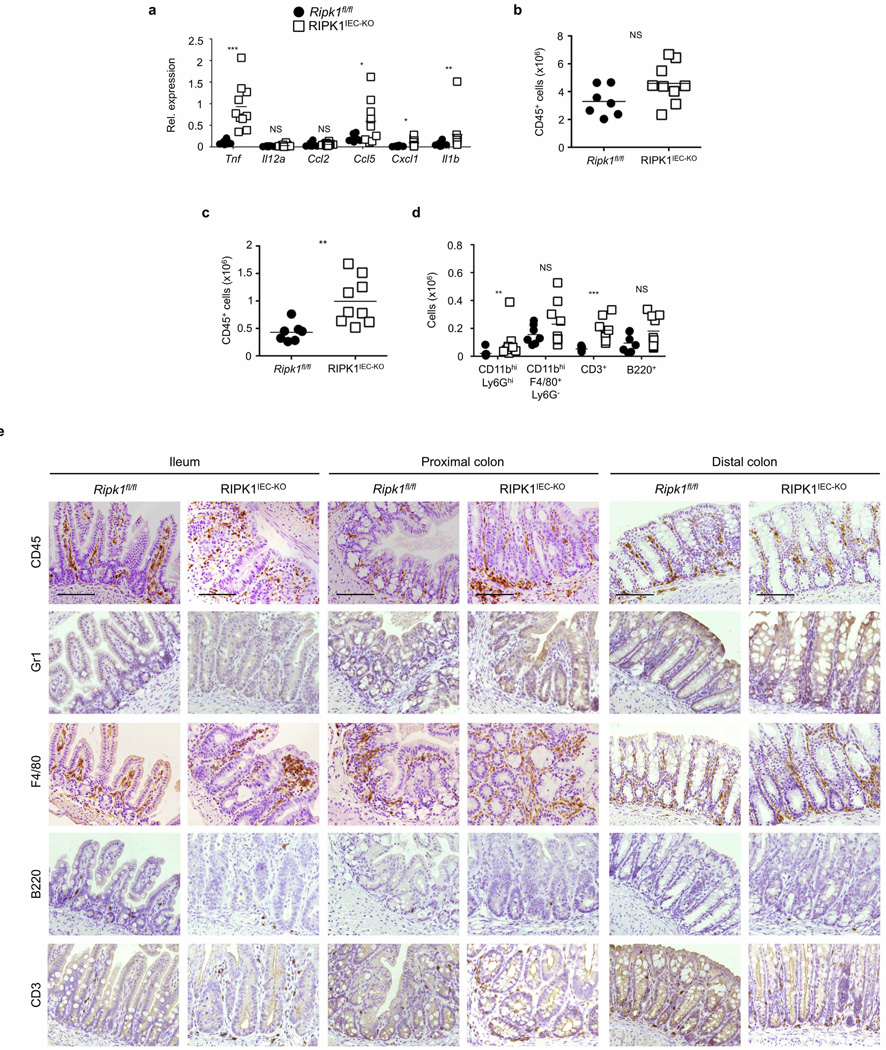
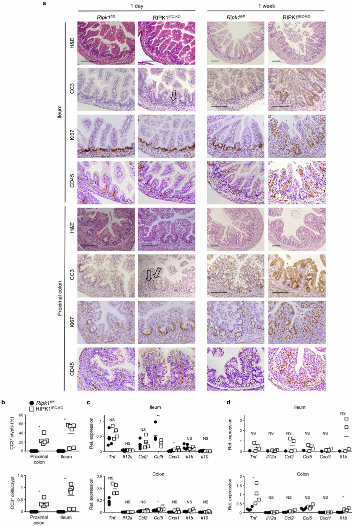
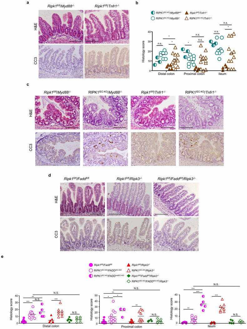
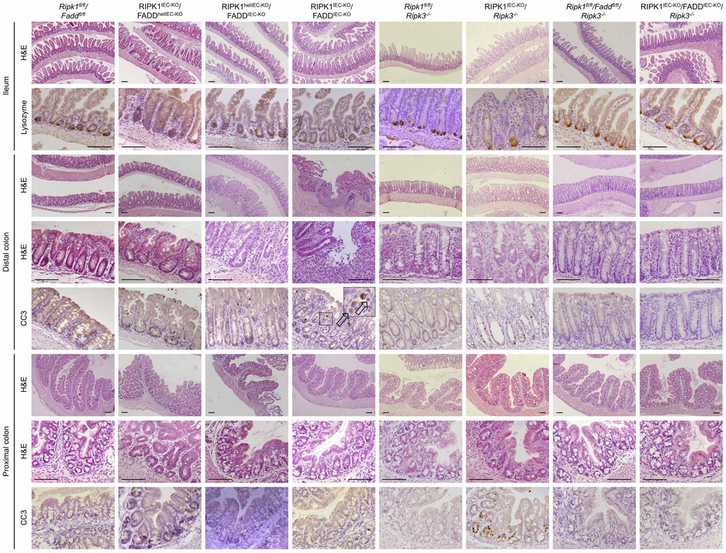
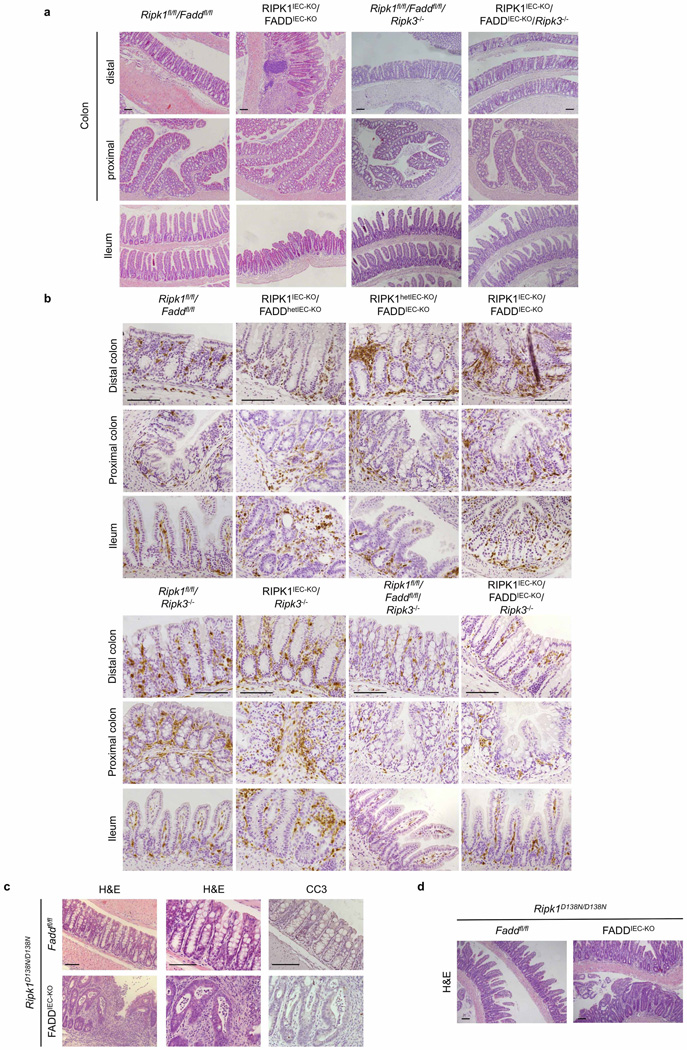
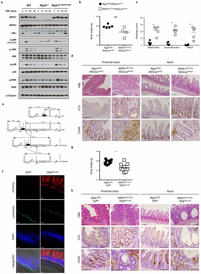
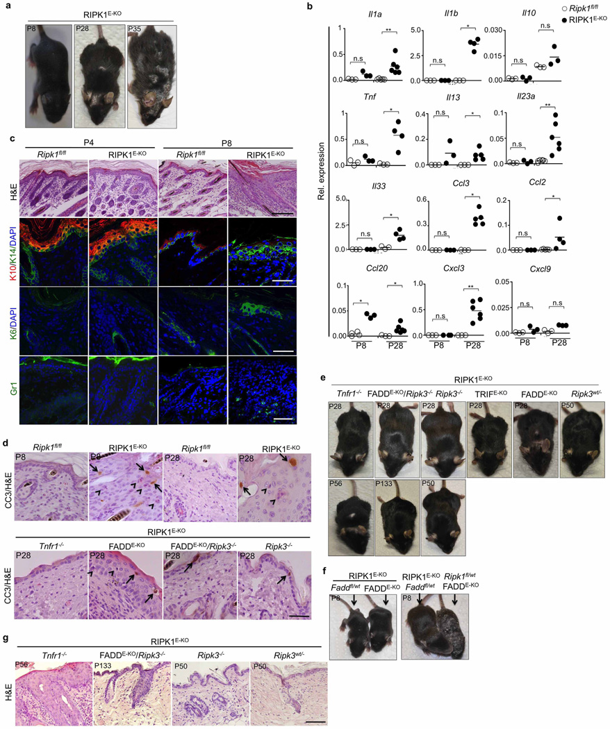
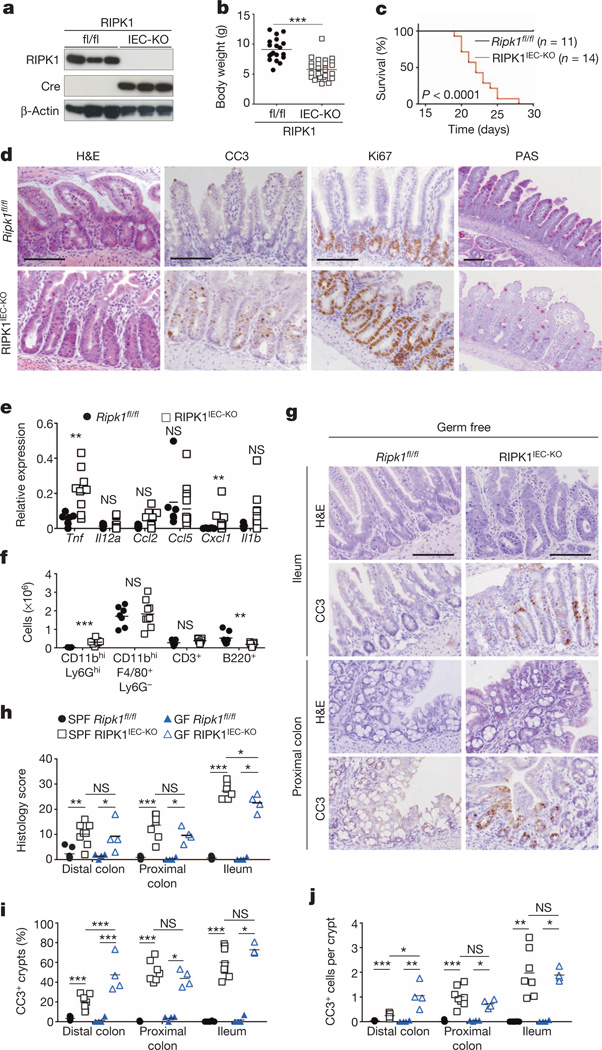
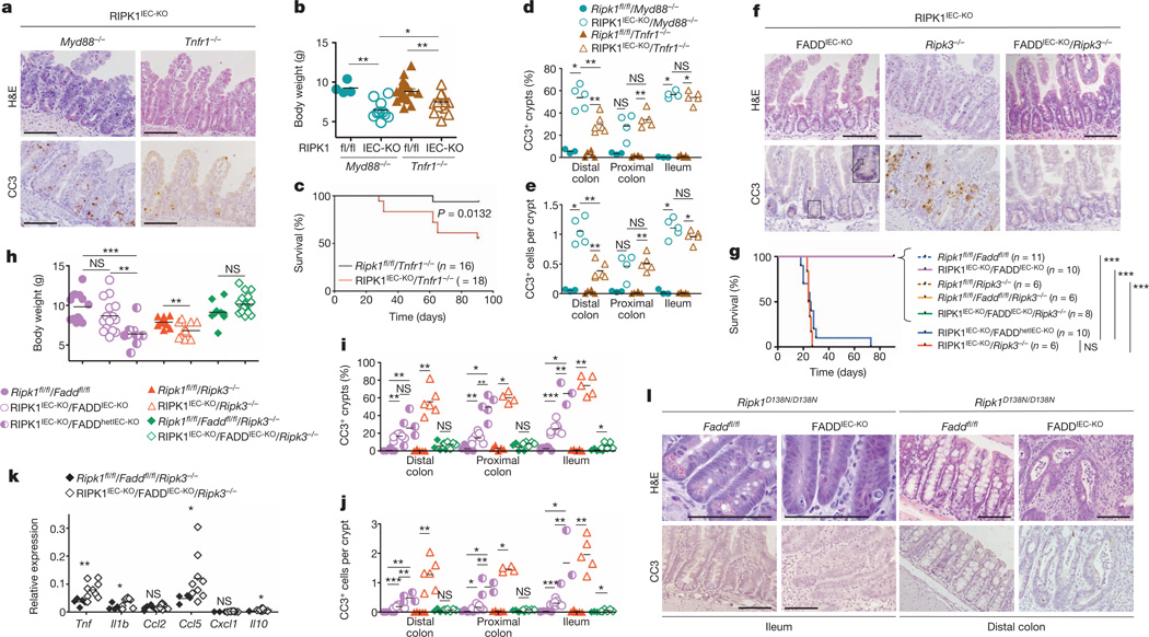
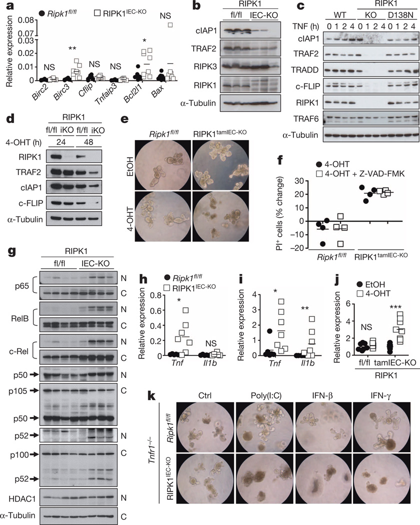
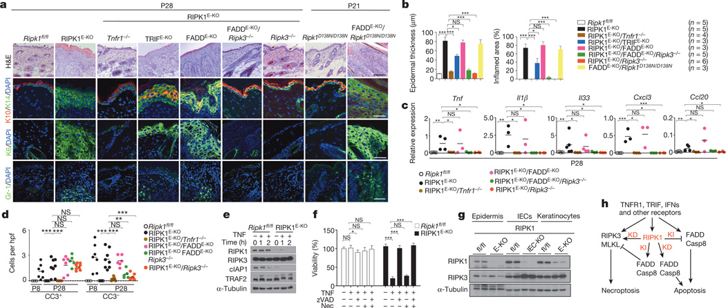
Necroptosis has emerged as an important pathway of programmed cell death in embryonic development, tissue homeostasis, immunity and inflammation. RIPK1 is implicated in inflammatory and cell death signalling and its kinase activity is believed to drive RIPK3-mediated necroptosis. Here we show that kinase-independent scaffolding RIPK1 functions regulate homeostasis and prevent inflammation in barrier tissues by inhibiting epithelial cell apoptosis and necroptosis. Intestinal epithelial cell (IEC)-specific RIPK1 knockout caused IEC apoptosis, villus atrophy, loss of goblet and Paneth cells and premature death in mice. This pathology developed independently of the microbiota and of MyD88 signalling but was partly rescued by TNFR1 (also known as TNFRSF1A) deficiency. Epithelial FADD ablation inhibited IEC apoptosis and prevented the premature death of mice with IEC-specific RIPK1 knockout. However, mice lacking both RIPK1 and FADD in IECs displayed RIPK3-dependent IEC necroptosis, Paneth cell loss and focal erosive inflammatory lesions in the colon. Moreover, a RIPK1 kinase inactive knock-in delayed but did not prevent inflammation caused by FADD deficiency in IECs or keratinocytes, showing that RIPK3-dependent necroptosis of FADD-deficient epithelial cells only partly requires RIPK1 kinase activity. Epidermis-specific RIPK1 knockout triggered keratinocyte apoptosis and necroptosis and caused severe skin inflammation that was prevented by RIPK3 but not FADD deficiency. These findings revealed that RIPK1 inhibits RIPK3-mediated necroptosis in keratinocytes in vivo and identified necroptosis as a more potent trigger of inflammation compared with apoptosis. Therefore, RIPK1 is a master regulator of epithelial cell survival, homeostasis and inflammation in the intestine and the skin.


Figures

Extended Data Figure 1
Extended Data Figure 1. Intestinal pathology in Ripk1−/− and RIPK1IEC-KO mice
a, Representative images of colonic and ileal sections from newborn Ripk1+/+ and Ripk1−/− mice stained with H&E or immunostained for CC3. b, Targeting strategy for the generation of mice with loxP-flanked (floxed (fl)) Ripk1 alleles. Exon 3 of the Ripk1 gene was flanked with loxP sites and an FRT-flanked neomycin (Neo) selectable cassette was introduced after the 3′ loxP site. The Neo cassette was excised by crossing the Ripk1neoFloxed mice with Flp-deleter mice. c, Representative Southern blots depicting the identification of correctly targeted embryonic stem (ES) cell clones by using 5′ and 3′ external probes. Arrows indicate a correctly targeted ES cell clone. Screening for the loxP site in intron 2 was performed by PCR (data not shown). Double combs allowing loading samples at two levels were used to maximize loading capacity of the gels for screening ES clones in 96-well plates. d, Immunoblot of colon IEC protein extracts from Ripk1fl/fl and RIPK1IEC-KO mice. e, f, Representative images of ileal (e) or colon (f) sections from Ripk1fl/fl and RIPK1IEC-KO mice stained with H&E, PAS or alkaline phosphatase (AP), or immunostained against lysozyme, CC3 or Ki67. Scale bars, 100 µm.
Extended Data Figure 2
Extended Data Figure 2. Mild intestinal inflammation in RIPK1IEC-KO mice
a, qRT–PCR analysis of cytokine and chemokine expression in colon tissue from Ripk1fl/fl and RIPK1IEC-KO mice. bd, FACS analysis of lamina propria leukocytes in the small intestine (b) and colon (c, d) of Ripk1fl/fl and RIPK1IEC-KO mice. e, Representative images from intestinal sections from Ripk1fl/fl and RIPK1IEC-KO mice immunostained with the indicated antibodies. Scale bars, 100 µm. *P ≤ 0.05, **P ≤ 0.01, ***P ≤ 0.005; NS, not significant.
Extended Data Figure 3
Extended Data Figure 3. Assessment of intestinal pathology in newborn and 1-week-old RIPK1IEC-KO mice
a, Representative images of intestinal sections from Ripk1fl/fl and RIPK1IEC-KO mice stained with H&E or immunostained with the indicated antibodies. Arrows indicate sparse CC3+ IECs in sections from 1-day-old animals. Scale bars, 100 µm. b, Quantification of crypts containing CC3+ cells and the number of CC3+ cells per crypt in intestinal sections of 1-week-old Ripk1fl/fl and RIPK1IEC-KO mice. c, d, qRT–PCR analysis of cytokine and chemokine expression in small intestinal and colon tissue from 1-day-old (c) and 1-week-old (d) Ripk1fl/fl and RIPK1IEC-KO mice. *P ≤ 0.05, **P ≤ 0.01, ***P ≤ 0.005; NS, not significant.
Extended Data Figure 4
Extended Data Figure 4. Antibiotic treatment does not prevent intestinal pathology in RIPK1IEC-KO and RIPK1tamIEC-KO mice
a, Experimental outline of tamoxifen injections (TAM; 1 mg, intraperitoneally). b, Immunoblot of small intestine IEC protein extracts from tamoxifen-treated Ripk1fl/fl and RIPK1tamIEC-KO mice. ce, Body weight change (c), Kaplan–Meier survival curve (d) and representative images of H&E- and CC3-stained intestinal sections (e) of tamoxifen-treated Ripk1fl/fl and RIPK1tamIEC-KO mice receiving antibiotics (+AB) or normal drinking water starting 4 weeks before tamoxifen administration. f, g, Body weight change (f) and representative images of H&E- and CC3-stained intestinal sections (g) in vehicle-injected Ripk1fl/fl and RIPK1tamIEC-KO mice. h, i, Body weight changes (h) and representative images of H&E-stained intestinal sections (i) of tamoxifen-injected Villin-CreERT2 mice (n = 3). jl, Body weight (j), Kaplan–Meier survival curve (k) and representative images of H&E- and CC3-stained intestinal sections (l) of Ripk1fl/fl and RIPK1IEC-KO mice treated with antibiotics from E17.5 to 3 weeks of age. Scale bars, 100 µm. Error bars represent mean values ± s.d. *P ≤ 0.05, **P ≤ 0.01, ***P ≤ 0.005; NS, not significant.
Extended Data Figure 5
Extended Data Figure 5. Role of MyD88, TNFR1, FADD and RIPK3 in the intestinal pathology of RIPK1IEC-KO mice
a, Representative images of ileal sections from the indicated mice as controls for the mice shown in Fig. 2a. b, Quantification of histological pathology score of the indicated mice. c, Representative images of H&E- or CC3-stained colon sections from the indicated mice. d, Representative images of H&E- or CC3-stained ileal sections from the indicated mice. Scale bars, 100 µm. e, Quantification of histological pathology score of the indicated mice. *P ≤ 0.05, **P ≤ 0.01, ***P ≤ 0.005; NS, not significant.
Extended Data Figure 6
Extended Data Figure 6. Epithelial-specific FADD deficiency reduces IEC apoptosis, ameliorates crypt atrophy but triggers erosive lesions in the colon of RIPK1IEC-KO mice
Representative images of ileal and colonic sections from the indicated mice stained with H&E or immunostained against lysozyme or CC3. Scale bars, 100 µm.
Extended Data Figure 7
Extended Data Figure 7. FADD and RIPK3 deficiency restores intestinal homeostasis in RIPK1IEC-KO mice and IEC necroptosis in FADDIEC-KO mice occurs in the absence of RIPK1 kinase activity
a, Representative images of H&E-stained colonic and ileal sections of adult mice with the indicated genotypes. b, Representative images of colonic and ileal sections of mice with the indicated genotypes immunostained for CD45. c, d, Representative images of colonic (c) and ileal (d) sections of adult Faddfl/fl/Ripk1D138N/D138N and FADDIEC-KO/Ripk1D138N/D138N mice stained with H&E or immunostained against CC3. Scale bars, 100 µm.
Extended Data Figure 8
Extended Data Figure 8. Assessment of the role of NF-κB and TRIF signalling in RIPK1IEC-KO mice
a, Immunoblot analysis of protein extracts from wild-type (WT), Ripk1−/− and Ripk1D138N/D138N MEFs after stimulation with 10 ng ml−1 recombinant murine TNF for the indicated time points. Data are representative of five independent experiments. bd, Body weight (b), quantification of histological pathology score (c) and representative H&E-, CC3- or CD45-stained intestinal sections (d) of mice with the indicated genotypes. e, Targeting strategy used for the generation of Triffl/fl mice. Exon 2 of the Trif gene was flanked with loxP sites. FRT-flanked neo and a stop cassette were placed upstream of the 3′ loxP site. A splice acceptor (SA)-mCherry cassette was introduced downstream of the 3′ loxP site. Cre-mediated recombination removes the Trif coding sequences and the stop cassette inducing the expression of mCherry by the Trif locus. The FRT-flanked neo was excised by crossing TrifneoFloxed mice with Flp-Deleter mice. f, Confocal microscopy images of near native small intestinal sections of Triffl/fl and TRIFIEC-KO mice stained with anti-lysozyme and DAPI. g, h, Body weight (g) and representative images of H&E-, CC3- or CD45-stained intestinal sections (h) of the indicated mice. Scale bars, 100 µm. *P ≤ 0.05, **P ≤ 0.01, ***P ≤ 0.005; NS, not significant.
Extended Data Figure 9
Extended Data Figure 9. Skin inflammation in RIPK1E-KO mice is dependent on RIPK3-mediated necroptosis
a, Representative macroscopic images of RIPK1E-KO mice. b, qRT–PCR analysis of pro-inflammatory cytokines and chemokines on total skin mRNA from Ripk1fl/fl and RIPK1E-KO mice. c, d, Representative images of skin sections from the indicated mice stained as indicated. In d, arrows point to CC3+ cells and arrowheads depict CC3 dying cells identified by their pyknotic nuclei and eosinophilic cytoplasm. Scale bars, 100 µm in H&E stained; 50 µm in immunostained sections. eg, Representative macroscopic pictures (e, f) and H&E-stained skin sections (g) of the indicated mice. Scale bars, 100 µm. *P ≤ 0.05, **P ≤ 0.01, ***P ≤ 0.005; NS, not significant.
Extended Data Figure 10
Extended Data Figure 10. CRISPR/Cas9-mediated knockout of MLKL prevents skin inflammation in RIPK1E-KO mice
a, Schematic depiction of the Mlkl locus. The sequence targeted by the small guide RNAs (sgRNA and truncated TRUNC_sgRNA) is indicated by capital letters. The position of the ATG and the binding sites of the primers used for genotyping and sequencing are indicated. The sgRNAs were designed to target a sequence containing an ApaLI restriction site that is used for RFLP analysis (underlined). The protospacer-adjacent motif (PAM) sequence is depicted in red. b, Sequences of the wild-type (WT) Mlkl locus and of the targeted Mlkl alleles of the two obtained RIPK1E-KO/Mlkl−/− mice. Mouse #150 carries one allele with one base pair (bp) deletion and one allele with one bp insertion. Mouse #374 carries one allele with one bp insertion and one allele with two bp insertions. All mutations cause frame shift and ablate MLKL protein expression. Mouse #150 was obtained using the full-length MLKL-sgRNA and mouse #374 was obtained using the truncated MLKL_sgRNA_TRUNC. c, Macroscopic appearance and histological images of skin sections from RIPK1E-KO/Mlkl−/− mouse #150 at the age of P95. Scale bars, 100 µm (H&E); 50µm (keratins and CC3).
Figure 1
Figure 1. Epithelial RIPK1 ablation causes microbiota-independent intestinal pathology
a, Immunoblot of small intestine IECs from Ripk1fl/fl (fl/fl) and RIPK1IEC-KO (IEC-KO) mice. b, c, Body weight (b) and Kaplan–Meier survival curve (c) of Ripk1fl/fl and RIPK1IEC-KO mice. d, Representative images of ileal sections from Ripk1fl/fl and RIPK1IEC-KO mice stained as indicated. H&E, haematoxylin and eosin. e, f, Quantitative polymerase chain reaction with reverse transcription (qRT–PCR) of cytokine and chemokine expression (e) and fluorescence-activated cell sorting (FACS) of lamina propria leukocytes (f) in the small intestine of Ripk1fl/fl and RIPK1IEC-KO mice. g, Representative images of H&E- or CC3-stained intestinal sections from germ-free Ripk1fl/fl and RIPK1IEC-KO mice. hj, Quantification of histological pathology score (h), crypts containing CC3+ cells (i) and CC3+ cells per crypt (j) in intestinal sections of the indicated mice. GF, germ free; SPF, specific pathogen free. Scale bars, 100 µm. *P ≤ 0.05, **P ≤ 0.01, ***P ≤ 0.005; NS, not significant.
Figure 2
Figure 2. Death of RIPK1-deficient IECs depends on FADD and RIPK3
a, Representative images of H&E- or CC3-stained ileal sections from the indicated mice. b, c, Body weight (b) and Kaplan–Meier survival curve (c) of the indicated mice. d, e, Quantification of crypts containing CC3+ cells (d) and CC3+ cell numbers per crypt (e) in intestinal sections of the indicated mice. f, Representative images of ileal sections from the indicated mice. Arrow depicts CC3 dying IEC. g, h, Kaplan–Meier survival curve (g) and body weight (h) of mice with the indicated genotypes. i, j, Quantification of crypts containing CC3+ cells (i) and CC3+ cell numbers per crypt (j) in intestinal sections of the indicated mice. k, qRT–PCR analysis of cytokine and chemokine expression in the small intestine of the indicated mice. l, Representative images of intestinal sections from 4-month-old mice with the indicated genotypes. Scale bars, 100 µm. *P ≤ 0.05, **P ≤ 0.01, ***P ≤ 0.005; NS, not significant.
Figure 3
Figure 3. RIPK1 prevents the degradation of pro-survival proteins
a, b, qRT–PCR of pro-survival genes (a) and immunoblot of the indicated proteins (b) in small intestine IECs from Ripk1fl/fl (fl/fl) and RIPK1IEC-KO (IEC-KO) mice. c, Immunoblot of wild-type (WT), Ripk1−/− (KO) and Ripk1D138N/D138N (D138N) MEFs stimulated with 10 ng ml−1 recombinant murine TNF. d, Immunoblot of small intestine organoids from Ripk1fl/fl (fl/fl) and RIPK1tamIEC-KO (iKO) mice treated with 4-hydroxytamoxifen (4-OHT) for 24 or 48 h. e, Ripk1fl/fl and RIPK1tamIEC-KO organoids were treated with vehicle (ethanol; EtOH) or 4-OHT for 20 h and photographed 48 h later. Original magnification, ×400. f, FACS quantification of propidium iodide (PI)+ cells in organoids 43 h after treatment with 4-OHT in the presence or absence of z-VAD-FMK. g, Immunoblot of cytoplasmic (C) and nuclear (N) proteins from small intestine IECs of four Ripk1fl/fl and four RIPK1IEC-KO mice with the indicated antibodies. h, i, qRT–PCR of Tnf and Il1b expression in IECs from small intestine (h) or colon (i) of Ripk1fl/fl and RIPK1IEC-KO mice. j, qRT–PCR of Tnf expression in small intestine organoids from Ripk1fl/fl and RIPK1IEC-KO mice 30 h after treatment with EtOH or 4-OHT. k, Representative images of Ripk1fl/fl/Tnfr1−/− and RIPK1IEC-KO/Tnfr1−/− organoids 16 h after treatment with 100 µg ml−1 poly(I:C), 1,000 U ml−1 IFN-β and 1,000 U ml−1 IFN-γ. Original magnification, ×400. Ctrl, control. Representative data are shown of four (e, f, k), three (c) or two (d, j) independent experiments. *P ≤ 0.05, **P ≤ 0.01, ***P ≤ 0.005; NS, not significant.
Figure 4
Figure 4. RIPK1 ablation causes keratinocyte necroptosis and skin inflammation
a, Representative images of skin sections from the indicated mice stained with H&E or the indicated antibodies. Nuclei stained with 4′,6-diamidino-2-phenylindole (DAPI). Scale bars, 100 µm (H&E); 50 µm (immunostainings). b, Microscopic quantification of epidermal thickness and inflamed skin area in the indicated mice. Error bars represent mean values ± standard error of the mean (s.e.m.). c, qRT–PCR of cytokine and chemokine expression in total skin from the indicated mice. d, Quantification of CC3+ and CC3 dying cells per high power field (hpf) in skin sections from the indicated mice. e, Immunoblot of primary keratinocytes stimulated with 20 ng ml−1 recombinant murine TNF for the indicated time points. f, Quantification of cell viability in primary keratinocytes treated with 50 ng ml−1 recombinant murine TNF in the presence or absence of z-VAD-FMK (zVAD; 20 µM) and necrostatin-1 (Nec; 30 µM). Mean values ± s.e.m. from biological triplicates (n = 3) are shown. g, Immunoblot of protein extracts from P4 epidermis, IECs or keratinocytes from mice of the indicated genotypes. h, Schematic model of the kinase-dependent (KD) and -independent (KI) functions of RIPK1 in apoptosis and necroptosis. Representative data of three independent experiments are shown in e, f. *P ≤ 0.05, **P ≤ 0.01, ***P ≤ 0.005; NS, not significant.

Comment in

References

    1. Oberst A, et al. Catalytic activity of the caspase-8–FLIPL complex inhibits RIPK3-dependent necrosis. Nature. 2011;471:363–367. - PMC - PubMed
    1. Kaiser WJ, et al. RIP3 mediates the embryonic lethality of caspase-8-deficient mice. Nature. 2011;471:368–372. - PMC - PubMed
    1. Zhang H, et al. Functional complementation between FADD and RIP1 in embryos and lymphocytes. Nature. 2011;471:373–376. - PMC - PubMed
    1. Bonnet MC, et al. The adaptor protein FADD protects epidermal keratinocytes from necroptosis in vivo and prevents skin inflammation. Immunity. 2011;35:572–582. - PubMed
    1. Welz PS, et al. FADD prevents RIP3-mediated epithelial cell necrosis and chronic intestinal inflammation. Nature. 2011;477:330–334. - PubMed

Publication types

MeSH terms

Substances