Diagnostic accuracy of endoscopic ultrasonography for rectal neuroendocrine neoplasms
- PMID: 25132764
- PMCID: PMC4130855
- DOI: 10.3748/wjg.v20.i30.10470
Diagnostic accuracy of endoscopic ultrasonography for rectal neuroendocrine neoplasms
Abstract
Aim: To investigate the diagnostic accuracy of endoscopic ultrasonography (EUS) for rectal neuroendocrine neoplasms (NENs) and the differential diagnosis of rectal NENs from other subepithelial lesions (SELs).
Methods: The study group consisted of 36 consecutive patients with rectal NENs histopathologically diagnosed using biopsy and/or resected specimens. The control group consisted of 31 patients with homochronous rectal non-NEN SELs confirmed by pathology. Epithelial lesions such as cancer and adenoma were excluded from this study. One EUS expert blinded to the histological results reviewed the ultrasonic images. The size, original layer, echoic intensity and homogeneity of the lesions and the perifocal structures were investigated. The single EUS diagnosis recorded by the EUS expert was compared with the histological results.
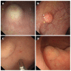
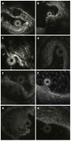
Results: All NENs were located at the rectum 2-10 cm from the anus and appeared as nodular (n = 12), round (n = 19) or egg-shaped (n = 5) lesions with a hypoechoic (n = 7) or intermediate (n = 29) echo pattern and a distinct border. Tumors ranged in size from 2.3 to 13.7 mm, with an average size of 6.8 mm. Homogeneous echogenicity was seen in all tumors except three. Apart from three patients (stage T2 in two and stage T3 in one), the tumors were located in the second and/or third wall layer without involvement of the fourth and fifth layers. In the patients with stage T1 disease, the tumors were located in the second wall layer only in seven cases, the third wall layer only in two cases, and both the second and third wall layers in 27 cases. Approximately 94.4% (34/36) of rectal NENs were diagnosed correctly by EUS, and 74.2% (23/31) of other rectal SELs were classified correctly as non-NENs. Eight cases of other SELs were misdiagnosed as NENs, including two cases of inflammatory lesions and one case each of gastrointestinal tumor, endometriosis, metastatic tumor, lymphoma, neurilemmoma, and hemangioma. The positive predictive value of EUS for rectal NENs was 80.9% (34/42), the negative predictive value was 92.0% (23/25), and the diagnostic accuracy was 85.1%.
Conclusion: EUS has satisfactory diagnostic accuracy for rectal NENs with good sensitivity, but unfavorable specificity, making the differential diagnosis of NENs from other SELs challenging.
Keywords: Diagnosis; Endoscopic ultrasonography; Neuroendocrine neoplasms; Rectum.
Figures


References
-
- Lepage C, Rachet B, Coleman MP. Survival from malignant digestive endocrine tumors in England and Wales: a population-based study. Gastroenterology. 2007;132:899–904. - PubMed
-
- Yao JC, Hassan M, Phan A, Dagohoy C, Leary C, Mares JE, Abdalla EK, Fleming JB, Vauthey JN, Rashid A, et al. One hundred years after “carcinoid”: epidemiology of and prognostic factors for neuroendocrine tumors in 35,825 cases in the United States. J Clin Oncol. 2008;26:3063–3072. - PubMed
-
- Niederle MB, Hackl M, Kaserer K, Niederle B. Gastroenteropancreatic neuroendocrine tumours: the current incidence and staging based on the WHO and European Neuroendocrine Tumour Society classification: an analysis based on prospectively collected parameters. Endocr Relat Cancer. 2010;17:909–918. - PubMed
-
- Garcia-Carbonero R, Capdevila J, Crespo-Herrero G, Díaz-Pérez JA, Martínez Del Prado MP, Alonso Orduña V, Sevilla-García I, Villabona-Artero C, Beguiristain-Gómez A, Llanos-Muñoz M, et al. Incidence, patterns of care and prognostic factors for outcome of gastroenteropancreatic neuroendocrine tumors (GEP-NETs): results from the National Cancer Registry of Spain (RGETNE) Ann Oncol. 2010;21:1794–1803. - PubMed
-
- Salazar R, Wiedenmann B, Rindi G, Ruszniewski P. ENETS 2011 Consensus Guidelines for the Management of Patients with Digestive Neuroendocrine Tumors: an update. Neuroendocrinology. 2012;95:71–73. - PubMed
Publication types
MeSH terms
LinkOut - more resources
Full Text Sources
Other Literature Sources

