doi: 10.5489/cuaj.1637.
Squamous cell carcinoma of the suprapubic tract: A rare presentation in patients with chronic indwelling urinary catheters
Affiliations
- PMID: 25132900
- PMCID: PMC4113586
- DOI: 10.5489/cuaj.1637
Item in Clipboard
Squamous cell carcinoma of the suprapubic tract: A rare presentation in patients with chronic indwelling urinary catheters
Can Urol Assoc J.
2014 Jul.
Abstract
Squamous cell carcinoma (SCC) of the bladder is uncommon, but can arise in the setting of long-term bladder catheterization and chronic inflammation. SCC can arise primarily from the suprapubic catheter tract, but fewer than 10 such cases have been reported. We document 2 cases of SCC arising from the suprapubic tract associated with chronic indwelling urinary catheters. SCC must be differentiated from granulomatous conditions, which are quite common in patients with suprapubic catheters.
Figures

A: Squamous cell carcinoma of the suprapubic tract in a 55-year-old male paraplegic patient; B: Close-up image of the fungating lesion.
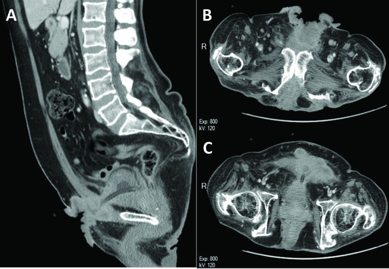
A computed tomography scan of a 55-year-old paraplegic patient with squamous cell carcinoma the suprapubic tract. A: Sagittal section demonstrating fungating lesion invading the rectus abdominus with extension to the anterior border of the pubic bone and dome of bladder. The suprapubic catheter and balloon can be observed. B–C: Transverse sections further delineating the extent of the tumour.

This well-differentiated tumour forms large irregularly shaped islands pushing deep into the underlying soft tissue (A). A high-power microscopic view shows that nuclear pleomorphism is minimal, mitoses are rare, and keratinization is evident (B).

Squamous cell carcinoma arising from the suprapubic catheter site in an 85-year-old female only 9 months after catheter placement. A: Appearance of lesion on initial presentation. B: Postoperative specimen including the skin lesion and subcutaneous tissues (top), suprapubic tract (middle), and partial cystectomy specimen (bottom).

Ulceration in the suprapubic tract with extensive involvement by infiltrating carcinoma. Necrosis is noted on the right (A). The tumour forms jagged sheets of pleomorphic cells and mitoses are frequent. Despite the aggressive cytological features, the presence of keratin pearls (arrows) is a clear evidence of squamous differentiation (B).
References
-
- Rous SN. Squamous cell carcinoma of the bladder. J Urol. 1978;120:561–2. - PubMed
-
- Kaufman JM, Fam B, Jacobs SC, et al. Bladder cancer and squamous metaplasia in spinal cord injury patients. J Urol. 1977;118:967–71. - PubMed
-
- Bejany DE, Lockhart JL, Rhamy RK. Malignant vesical tumors following spinal cord injury. J Urol. 1987;138:1390–2. - PubMed
LinkOut - more resources
Full Text Sources
Other Literature Sources
Research Materials