Review
. 2014 Aug 16;6(8):345-51.
doi: 10.4253/wjge.v6.i8.345.

Endoscopic retrograde cholangiopancreatography in patients with altered anatomy: How to deal with the challenges?

Affiliations
Review

Endoscopic retrograde cholangiopancreatography in patients with altered anatomy: How to deal with the challenges?

Tom G Moreels. World J Gastrointest Endosc. .

Abstract

Endoscopic retrograde cholangiopancreatography (ERCP) in patients with surgically altered anatomy is challenging. Several operative interventions of both the gastrointestinal tract and the biliary and/or pancreatic system lead to altered anatomy, rendering ERCP more difficult or even impossible with a conventional side-viewing duodenoscope. Adapted endoscopes are available to reach the biliopancreatic system and to perform ERCP in patients with altered anatomy. However, both technical difficulties and complications determine the procedure's success. Different technical approaches have been described and are highly dependent on local expertise and endoscopic equipment. Standardized practical guidelines are currently unavailable. This review focuses on the challenges encountered during ERCP in patients with altered anatomy and how to deal with them. The first challenge is reaching the papilla or the bilioenteric/pancreatoenteric anastomosis in the patient with postoperative altered anatomy. The second challenge is the cannulation of the biliopancreatic system and performing all conventional ERCP interventions and the third challenge is the control of possible complications. The available literature data on this topic is reviewed and illustrated with clinical cases.

Keywords: Altered anatomy; Billroth; Endoscopic retrograde cholangiopancreatography; Roux-en-Y.


Figures
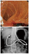
Figure 1
Figure 1
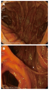
View Billroth II. A: Endoscopic view of Billroth II gastrojejunostomy with sharp angulation towards the afferent limb while retracting the single-balloon enteroscope. Note the mucosal tear at the short angle of the afferent limb at the end of the endoscopic retrograde cholangiopancreatography procedure; B: Radiological view of the looping position of the single-balloon enteroscope in the stomach of a patient with Billroth II partial gastrectomy. The tip of the enteroscope is located in the blind end of the duodenum. The white arrow denotes the position of the deflated overtube balloon.
Figure 2
Figure 2
Endoscopic view of Roux-en-Y reconstruction. A: Endoscopic view of an end-to-side Roux-en-Y reconstruction with view on the afferent limb (right) and common limb (left). The white arrow denotes the operative scar of the anastomosis; B: Endoscopic view of a side-to-side Roux-en-Y reconstruction with view on the afferent limb (middle), common limb (left) and blind ending limb (right). The white arrow denotes the operative scar of the anastomosis.
Figure 3
Figure 3
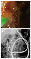
View. A: Endoscopic view of the distal approach to cannulate an intact papilla of Vater using a straight catheter with a forward-viewing endoscope. In order to cannulate in the direction of the common bile duct, the papilla is rotated into the 7 o’clock position; B: Radiological view of the double-balloon enteroscope in a patient with Roux-en-Y gastric bypass. The distal approach with a forward-viewing endoscope allows straight cannulation of the common bile duct (white arrow) in line with the direction of the working channel.
Figure 4
Figure 4
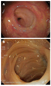
Endoscopic view of bilioenteric anastomosis. A: Endoscopic view of a normal end-to-side bilioenteric anastomosis; B: Endoscopic view of a stenosis at the level of the bilioenteric anastomosis. Only scar tissue (white arrow) indicates the location of the anastomosis without visible opening.
Figure 5
Figure 5
A patient with Roux-en-Y gastric bypass. A: Computed tomography of a retroperitoneal perforation (white arrow) at the level of papilla of Vater after sphincterotomy and sphincteroplasty in a patient with Roux-en-Y gastric bypass; B: Radiological view of hepatic capsule dehiscence without free abdominal air (white arrow) due to barotrauma in the closed afferent limb during single-balloon enteroscopy endoscopic retrograde cholangiopancreatography in a patient with Billroth II partial gastrectomy. Common bile duct stone retrieval with a basket is being performed.
Figure 6
Figure 6
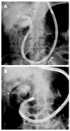
Radiological view. A: Radiological view of direct cholangioscopy with the single-balloon enteroscope inside the common bile duct and with the overtube (white arrow) inside the afferent limb of a Billroth II patient; B: Radiological view of free air around the contrast-filled gallbladder (white arrow) due to barotrauma during single-balloon enteroscopy endoscopic retrograde cholangiopancreatography in a Billroth II patient.

References

    1. Cotton PB. ERCP overview. A 30-year perspective. In: Advanced digestive endoscopy: ERCP., editor. Cotton P, Leung J, editors. Massachusetts: Blackwell Publishing Ltd; 2005. pp. 1–8.
    1. Dumonceau JM, Andriulli A, Deviere J, Mariani A, Rigaux J, Baron TH, Testoni PA. European Society of Gastrointestinal Endoscopy (ESGE) Guideline: prophylaxis of post-ERCP pancreatitis. Endoscopy. 2010;42:503–515. - PubMed
    1. Adler DG, Baron TH, Davila RE, Egan J, Hirota WK, Leighton JA, Qureshi W, Rajan E, Zuckerman MJ, Fanelli R, et al. ASGE guideline: the role of ERCP in diseases of the biliary tract and the pancreas. Gastrointest Endosc. 2005;62:1–8. - PubMed
    1. Moreels TG. ERCP in the patient with surgically altered anatomy. Curr Gastroenterol Rep. 2013;15:343. - PubMed
    1. Lee A, Shah JN. Endoscopic approach to the bile duct in the patient with surgically altered anatomy. Gastrointest Endosc Clin N Am. 2013;23:483–504. - PubMed