. 2014 Aug 15;6(8):289-300.
doi: 10.4251/wjgo.v6.i8.289.

In vitro effects of polyphenols on colorectal cancer cells

Affiliations

In vitro effects of polyphenols on colorectal cancer cells

Barbara Pampaloni et al. World J Gastrointest Oncol. .

Abstract

Aim: To investigate the effects of quercetin and genistein on colon cancer cell proliferation and their estrogen receptor β (ERβ) expression.

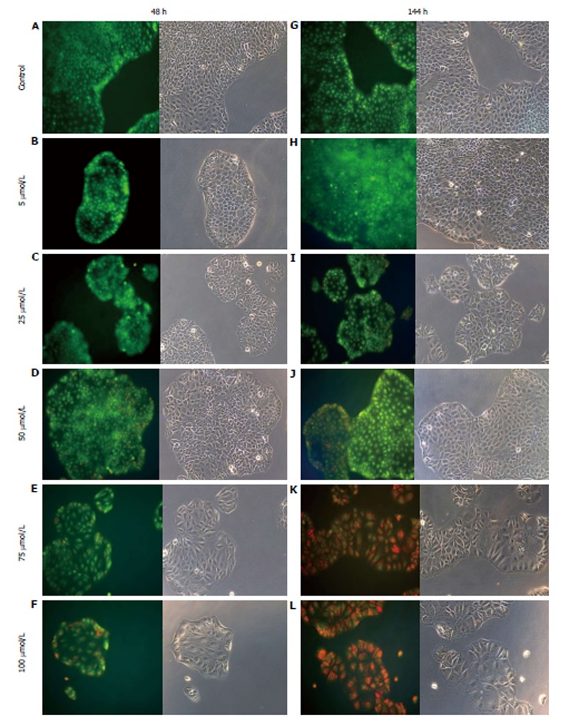
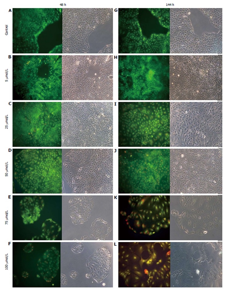
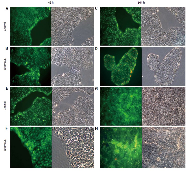
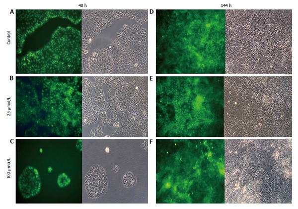
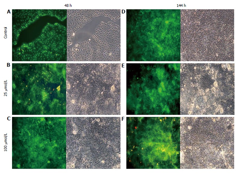
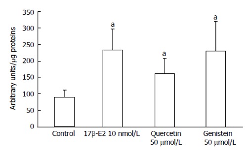
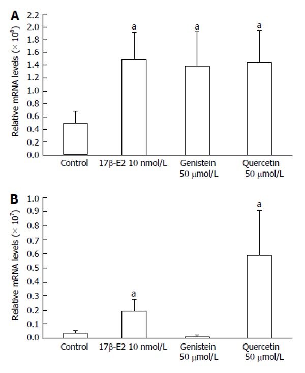
Methods: Colon cancer cells were stably transfected with a mammalian expression vector to overexpress ERβ (HCT8-β8-expressing cells) or a control vector (HCT8-pSV2neo-expressing cells). The proliferation of these cells was examined after treatment with quercetin or genistein (5-100 μmol/L), or 10 nmol/L 17β-estradiol (17β-E2). Cell viability was examined by acridine orange staining following treatments for 48 or 144 h. Effects of quercetin and genistein on ERβ transcriptional transactivation were examined by luciferase activity in HCT8-β8-expressing cells transiently transfected with a pEREtkLUC reporter vector. In addition, the regulation of ERβ transcription by phytoestrogens and 17β-E2 was examined by quantitative polymerase chain reaction.

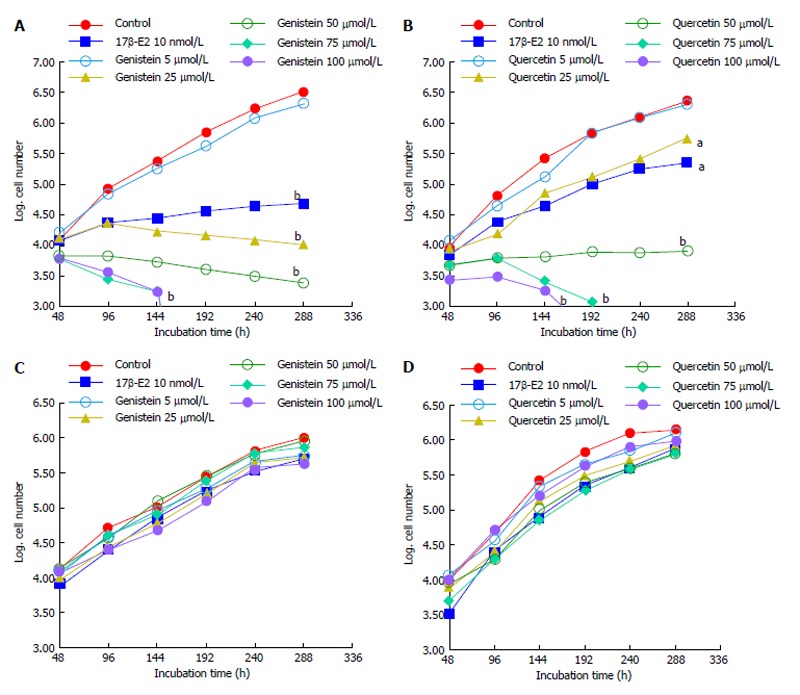
Results: Proliferation of HCT8-β8-expressing cells was not reduced low doses (5 μmol/L) of quercetin and genistein, while it was reduced at 25-50 μmol/L with an effect similar to 10 nmol/L 17β-E2. Treatment with doses of phytoestrogens ≥ 75 μmol/L completely blocked cell growth and reduced overall cell counts, however no effects at any dose were observed in HCT8-pSV2neo-expressing cells. These results were supported by viability staining that revealed acridine orange-stained lysosomes with high doses or extended treatment periods. Genistein and quercetin (50 μmol/L) significantly increased ER-responsive luciferase activity similar to 10 nmol/L 17β-E2 (P < 0.05). Furthermore, genistein and quercetin (50 μmol/L), as well as 10 nmol/L 17β-E2 significantly increased ERβ mRNA levels in HCT8-β8-expressing cells (P < 0.05). In addition, treatment of HCT8-pSV2neo-expressing cells with 50 µmol/L quercetin or 10 nmol/L 17β-E2 significantly increased ERβ mRNA levels compared to untreated controls (P < 0.05), though the absolute levels were much lower than in HCT8-β8-expressing cells.

Conclusion: The antitumorigenic effects of the phytoestrogenic compounds quercetin and genistein on colon cancers cells occur through ERβ activity and expression.

Keywords: Estrogen receptor; Genistein; HCT8-pSV2neo; HCT8-β8 cells; Quercetin.


Figures

Figure 1
Figure 1
Effects of polyphenols on cell growth. A: Growth of HCT8-â8-expressing cells in the presence of genistein and 17â-E2; B: Growth of HCT8-β8-expressing cells in the presence of quercetin and 17β-E2; C: Growth of HCT8-pSV2neo-expressing cells in the presence of genistein and 17β-E2; D: Growth of HCT8-pSV2neo-expressing cells in the presence of quercetin and 17β-E2. Values are the means of triplicates; aP < 0.05 vs control; bP < 0.01 vs control.
Figure 2
Figure 2
Treatment of HCT8-β8-expressing cells with genistein. HCT8-β8-expressing cells were treated with various concentrations of genistein for 48 h (A-F) or 144 h (G-L) and stained with acridine orange. Nuclei and mitochondria appear green, whereas lysosomes appear red-orange under fluorescence, adjacent to corresponding phase contrast images (magnification × 20).
Figure 3
Figure 3
Treatment of HCT8-β8-expressing cells with quercetin. HCT8- β8-expressing cells were treated with various concentrations of quercetin for 48 h (A-F) or 144 h (G-L) and stained with acridine orange. Nuclei and mitochondria appear green, whereas lysosomes appear red-orange under fluorescence, adjacent to corresponding phase contrast images (magnification, × 20).
Figure 4
Figure 4
Treatment of cells with 17β-E2. A-D: HCT8-β8-expressing cells; or E-H: HCT8-pSV2neo-expressing cells were treated with 10 nmol/L 17β-E2 for 48 h (A, B, E, F) or 144 h (C, D, G, H) and stained with acridine orange. Nuclei and mitochondria appear green, whereas lysosomes appear red-orange under fluorescence, adjacent to corresponding phase contrast images (magnification × 20).
Figure 5
Figure 5
Treatment of HCT8-pSV2neo-expressing cells with genistein. A-F: HCT8-pSV2neo-expressing cells were treated with 25 μmol/L (B and E) or 100 μmol/L (C and F) genistein for 48 h (A-C) or 144 h (D-F) and stained with acridine orange. Nuclei and mitochondria appear green, whereas lysosomes appear red-orange under fluorescence, adjacent to corresponding phase contrast images (magnification × 20).
Figure 6
Figure 6
Treatment of HCT8-pSV2neo-expressing cells with quercetin. A-F: HCT8-pSV2neo-expressing cells were treated with 25 μmol/L (B and E) or 100 μmol/L (C and F) quercetin for 48 h (A-C) or 144 h (D-F) and stained with acridine orange. Nuclei and mitochondria appear green, whereas lysosomes appear red-orange under fluorescence, adjacent to corresponding phase contrast images (magnification × 20).
Figure 7
Figure 7
Induction of EREtkLUC reporter gene activity. Treatment of HCT8-â8-expressing cells with 17â-E2, genistein and quercetin induces EREtk expression observed as relative luciferase activity. Values are the mean ± SD of triplicates; aP < 0.05 vs control.
Figure 8
Figure 8
Expression of ERβ mRNA levels by quantitative real-time reverse transcription-polymerase chain reaction. Induction of ERβ expression by 17β-E2, genistein and quercetin in A: HCT8-β8-expressing cells; B: HCT8-pSV2neo-expressing cells. The results are expressed relative to RPS18 mRNA levels. Values are the mean ± SD of quadruplicates; aP < 0.05 vs control.

References

    1. American Cancer Society. Cancer Facts and Figures 2010. Atlanta: American Cancer Society; 2010. Available from: http: //www.cancer.org/acs/groups/content/@nho/documents/document/acspc-....
    1. Chen L, Crawford JM. Tratto Gastrointestinale. In: Robbins and Cotran., editor. Pathologic Basis of Disease, 7th ed. Milan: Elsevier; 2006. pp. 797–877.
    1. Wei EK, Colditz GA, Giovannucci EL, Fuchs CS, Rosner BA. Cumulative risk of colon cancer up to age 70 years by risk factor status using data from the Nurses’ Health Study. Am J Epidemiol. 2009;170:863–872. - PMC - PubMed
    1. Lacassagne A. Endocrine factors concerned in the genesis of experimental mammary carcinoma. J Endocrinol. 1955;13:ix–xviii. - PubMed
    1. American Cancer Society. Cancer Facts and Figures 2007. Atlanta: American Cancer Society; 2007. Available from: http: //www.cancer.org/acs/groups/content/@nho/documents/document/caff20....

LinkOut - more resources