. 2014 Aug;6(8):1150-8.
doi: 10.3978/j.issn.2072-1439.2014.05.11.

Heart-lung transplantation

Affiliations

Heart-lung transplantation

Charles B Huddleston et al. J Thorac Dis. 2014 Aug.

Abstract

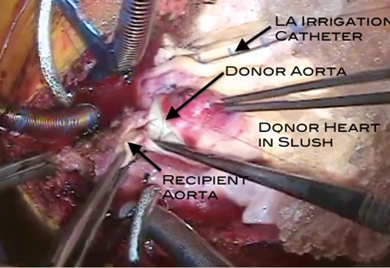
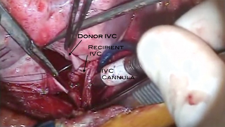
Heart-lung transplantation itself is not a particularly difficult operation technically. It is the setting in which this procedure is performed which is difficult. The three issues of importance in a successful outcome are appropriate harvest of the heart-lung bloc from the donor, careful explant of the heart and lungs of the recipient, and finally the implant of the heart-lung bloc into the recipient. None of this requires extraordinary technical skill, but does require careful coordination and planning as well as adhering to some fundamental principles. One of the major pitfalls encountered is bleeding related to the explant procedure. Another is graft failure related to harvest and/or the implant procedure. The third is injury to either the phrenic nerve(s) or the left recurrent laryngeal nerve related to the explant procedure. Heart-lung transplantation is a major investment in resources of all sorts including financial, personnel, as well as the organs themselves. It is absolutely imperative that this procedure be performed only by experienced surgeons in centers with established expertise.

Keywords: Technique; heart-lung transplantation; transplantation.


Figures

Figure 1
Figure 1
Preparation of the heart-lung bloc. The organs are taken from the cold storage and brought up onto the operative field. The lungs and heart should remain in a slush solution as much as possible during this preparation. All the excess pericardial tissue is removed followed by the esophagus and aortic tissue taken with the organs at the time of the harvest. The trachea is identified and transected at a point approximately 1-2 cartilaginous rings above the takeoff of the right upper lobe bronchus. There is always an impressive collection of mucoid secretions present. These are cultured and then suctioned completely to remove as much as possible (8).
Figure 2
Figure 2
(A) The stapled esophagus is removed by dissecting it away from its mediastinal attachments. The remaining pericardium and aorta have been trimmed away. The trachea is divided above the bifurcation; (B) The trachea is divided above the bifurcation and the staple line is removed; (C) The left atrial appendage which had been amputated is closed with a pursestring to allow the insertion of a catheter for irrigating the left side cardiac structures with cold crystalloid solution.
Figure 3
Figure 3
Recipient cardiectomy. The operation is performed via a midline sternotomy. After dissection of as much of the heart and lungs as possible off bypass, bicaval/aortic cannulation is performed and the patient is placed on cardiopulmonary bypass. The aorta is cross-clamped. The caval tapes are snared. The right atrium is opened in the midportion of the anterior wall. There is a tremendous amount of pulmonary venous return in this case because of an extensive aorto-pulmonary collateral network related to the longstanding cyanosis. The aorta is divided. The atrial incision is taken superiorly around the right atrial appendage and across the atrial septum to the roof of the left atrium. The incision inferiorly is taken toward the coronary sinus. There is no pulmonary artery connection in this patient so that is not divided. The remaining atrial wall holding the heart in is divided and the heart removed from the operative field. This leaves behind the atrial mass (left and right atrial tissue) (9).
Figure 4
Figure 4
Left pneumonectomy. Bilateral pneumonectomies are performed. This video demonstrates highlights from the left pneumonectomy. Adhesions along the pleural surface and mediastinum are taken down with the electrocautery. The inferior pulmonary ligament is divided with the electrocautery and this is further used to go through the pulmonary veins and arteries, rather than taking the time to ligate these vessels; the veins are already open into the pericardium and the only flow into the arteries is via aortopulmonary collateral circulation. The bronchus is dissected free. A stapling device is then applied to the bronchus and it is divided distally. The lung should be able to be removed at this point. A similar procedure is performed for the right lung (10).
Figure 5
Figure 5
Preparation of the IVC and SVC. The remaining atrial tissue is removed. This transplant will be performed using caval anastomoses (rather than a right atrial anastomosis), so as the atrial tissue is removed there should be a sufficient cuff left behind to which the donor SVC and IVC will be sewn. The orifices of the pulmonary veins are easily visible. Liberal use of the electrocautery is evident. Once this tissue is removed, the chest is devoid of the heart and lungs, leaving behind an impressive cavity (11).
Figure 6
Figure 6
(A) This demonstrates the preparation of the SVC cuff. The excess right atrial tissue is trimmed leaving a sufficient rim for the anastomosis to the donor SVC; (B) Similarly for the IVC a cuff of the right atrium should be left attached to the IVC to allow sufficient tissue and length for the IVC anastomosis to the donor heart. SVC, superior vena cava; IVC, inferior vena cava.
Figure 7
Figure 7
Removal of bronchi and distal trachea. The remaining mainstem bronchi and distal trachea are now excised. The mainstem bronchi are grasped with Allis clamps to assist with the dissection. Placing a stay suture on the more proximal trachea is often helpful for exposure during the tracheal anastomosis. Avoid dissection along the lateral portion of the trachea as much as possible to maintain adequate blood supply to this area. The trachea is divided as distally as possible, remaining cephalad to the mainstem bronchi. Once the anterior wall of the trachea is incised it is obvious from the bleeding that the blood supply is excellent. The remaining portion of the trachea is incised and the distal segment is dissected away from the mediastinum, leaving an open trachea in the mediastinum prepared for the anastomosis (12).
Figure 8
Figure 8
With both lungs and the heart out of the chest the only things remaining are pericardium on both sides to include the phrenic nerves and mediastinal tissue with the lymphatics, esophagus and descending thoracic aorta. Note the large opening posterior to each leaf of pericardium posteriorly to allow the passage of the donor lungs of the heart-lung bloc into the respective thoracic cavities.
Figure 9
Figure 9
Placement of the heart-lung bloc. A pathway has been created posterior to the phrenic nerves on each side. The heart-lung bloc is lowered into the chest passing one lung (the left in this case) into its thoracic space and then the other. Because the lungs remain somewhat inflated, this may require some gentle encouragement to get each lung into its respective position. The heart should be well aligned once this is accomplished (13).
Figure 10
Figure 10
The tracheal anastomosis is the first connection for the heart-lung bloc. This can be done with either a running simple suture technique or using the running technique for the membranous portion and interrupted stitches for the cartilaginous portion of the trachea.
Figure 11
Figure 11
Tracheal anastomosis. This is usually done with a continuous suture of 4-0 polypropylene suture. Other monofilament absorbable suture is certainly reasonable to use as well. Once this is completed, the suture line should be covered with the paratracheal and lymphatic tissue nearby (14).
Figure 12
Figure 12
Placement of the LA catheter. Prior to performing the aortic anastomosis, a catheter is placed via the left atrial appendage into the body of the left atrium. A standard LV vent is appropriate with an attachment that allows for the instillation of cold crystalloid solution during the aortic anastomosis. This is placed at the site of the LS appendage amputation of the appendage performed at the time of the organ harvest. The infusion of cold crystalloid solution via this catheter keeps the heart cold, but also provides a means of air evacuation. There is no pulmonary venous return to the left atrium until there is antegrade flow through the lungs because the bronchial circulation has been divided. The fluid is run through this catheter at a rate that results in a low flow of fluid from the aorta (15).
Figure 13
Figure 13
The aortic anastomosis is performed here using a simple running suture technique.
Figure 14
Figure 14
Aortic anastomosis. This is a simple end-to-end connection. As this is being completed the cold crystalloid solution infusing via the LA catheter can be seen coming out of the open portion of the aortic anastomosis. This aids in the de-airing process. Once this is done the aortic cross-clamp can be removed to re-perfuse the heart (16).
Figure 15
Figure 15
The IVC anastomosis is performed in an end-to-end fashion. IVC, inferior vena cava.
Figure 16
Figure 16
IVC anastomosis. This is generally done with the cross-clamp off the aorta and the heart re-perfused. However, the coronary sinus return to the right atrium often obscures the anastomotic site. This connection is also a simple end-to-end anastomosis (17).
Figure 17
Figure 17
SVC anastomosis. Again, this is a simple end-to-end anastomosis. Care should be taken to avoid pursestringing this anastomosis by interrupting the suture line in three or four locations. If there is significant size discrepancy it is probably better to open the smaller vessel longitudinally and sew on a patch to enlarge this (18).
Figure 18
Figure 18
Functioning transplanted heart and lungs. This merely shows the heart and lungs once off cardiopulmonary bypass. The heart is contracting vigorously and the lungs appear appropriately pink (19).

References

    1. Webb WR, Howard HS. Cardio-pulmonary transplantation. Surg Forum 1957;8:313-7 - PubMed
    1. Lower RR, Stofer RC, Hurley EJ, et al. Complete homograft replacement of the heart and both lungs. Surgery 1961;50:842-5 - PubMed
    1. Nakae S, Webb WR, Theodorides T, et al. Respiratory function following cardiopulmonary denervation in dog, cat, and monkey. Surg Gynecol Obstet 1967;125:1285-92 - PubMed
    1. Reitz BA, Burton NA, Jamieson SW, et al. Heart and lung transplantation: autotransplantation and allotransplantation in primates with extended survival. J Thorac Cardiovasc Surg 1980;80:360-72 - PubMed
    1. Reitz BA, Wallwork JL, Hunt SA, et al. Heart-lung transplantation: successful therapy for patients with pulmonary vascular disease. N Engl J Med 1982;306:557-64 - PubMed