

Intra-QT spectral coherence as a possible noninvasive marker of sustained ventricular tachycardia
- PMID: 25133170
- PMCID: PMC4123476
- DOI: 10.1155/2014/583035
Intra-QT spectral coherence as a possible noninvasive marker of sustained ventricular tachycardia
Abstract
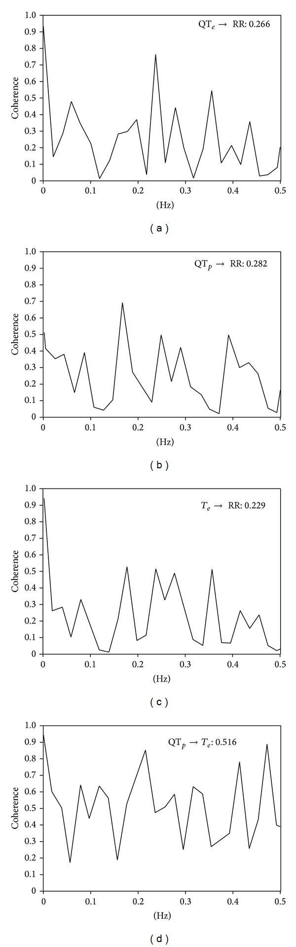
Sudden cardiac death is the main cause of mortality in patients affected by chronic heart failure (CHF) and with history of myocardial infarction. No study yet investigated the intra-QT phase spectral coherence as a possible tool in stratifying the arrhythmic susceptibility in patients at risk of sudden cardiac death (SCD). We, therefore, assessed possible difference in spectral coherence between the ECG segment extending from the q wave to the T wave peak (QTp) and the one from T wave peak to the T wave end (Te) between patients with and without Holter ECG-documented sustained ventricular tachycardia (VT). None of the QT variability indexes as well as most of the coherences and RR power spectral variables significantly differed between the two groups except for the QTp-Te spectral coherence. The latter was significantly lower in patients with sustained VT than in those without (0.508 ± 0.150 versus 0.607 ± 0.150, P < 0.05). Although the responsible mechanism remains conjectural, the QTp-Te spectral coherence holds promise as a noninvasive marker predicting malignant ventricular arrhythmias.
Figures
References
-
- Tereshchenko LG, Cygankiewicz I, McNitt S, et al. Predictive value of beat-to-beat qt variability index across the continuum of left ventricular dysfunction competing risks of noncardiac or cardiovascular death and sudden or nonsudden cardiac death. Circulation: Arrhythmia and Electrophysiology. 2012;5(4):719–727. - PMC - PubMed
-
- Berger RD, Kasper EK, Baughman KL, Marban E, Calkins H, Tomaselli GF. Beat-to-beat QT interval variability: Novel evidence for repolarization lability in ischemic and nonischemic dilated cardiomyopathy. Circulation. 1997;96(5):1557–1565. - PubMed
-
- Piccirillo G, Magrì D, Pappadà MA, et al. Autonomic nerve activity and the short-term variability of the Tpeak-Tend interval in dogs with pacing-induced heart failure. Heart Rhythm. 2012;9(12):2044–2050. - PubMed
MeSH terms
Substances
LinkOut - more resources
Full Text Sources
Other Literature Sources
