. 2014 Sep;124(9):3901-12.
doi: 10.1172/JCI73777. Epub 2014 Aug 18.

Cyclooxygenase-2-dependent lymphangiogenesis promotes nodal metastasis of postpartum breast cancer

Cyclooxygenase-2-dependent lymphangiogenesis promotes nodal metastasis of postpartum breast cancer

Traci R Lyons et al. J Clin Invest. 2014 Sep.

Abstract

Breast involution following pregnancy has been implicated in the high rates of metastasis observed in postpartum breast cancers; however, it is not clear how this remodeling process promotes metastasis. Here, we demonstrate that human postpartum breast cancers have increased peritumor lymphatic vessel density that correlates with increased frequency of lymph node metastases. Moreover, lymphatic vessel density was increased in normal postpartum breast tissue compared with tissue from nulliparous women. In rodents, mammary lymphangiogenesis was upregulated during weaning-induced mammary gland involution. Furthermore, breast cancer cells exposed to the involuting mammary microenvironment acquired prolymphangiogenic properties that contributed to peritumor lymphatic expansion, tumor size, invasion, and distant metastases. Finally, in rodent models of postpartum breast cancer, cyclooxygenase-2 (COX-2) inhibition during the involution window decreased normal mammary gland lymphangiogenesis, mammary tumor-associated lymphangiogenesis, tumor cell invasion into lymphatics, and metastasis. Our data indicate that physiologic COX-2-dependent lymphangiogenesis occurs in the postpartum mammary gland and suggest that tumors within this mammary microenvironment acquire enhanced prolymphangiogenic activity. Further, our results suggest that the prolymphangiogenic microenvironment of the postpartum mammary gland has potential as a target to inhibit metastasis and suggest that further study of the therapeutic efficacy of COX-2 inhibitors in postpartum breast cancer is warranted.


Figures

Figure 5
Figure 5. COX-2–dependent lymphangiogenesis, lymphatic vessel invasion, and lung metastasis in a xenograft model of postpartum breast cancer.
(A) shCOX-2 cells induce fewer branches in tube formation assays compared with shGFP MCF10DCIS (shGFP) vector controls. (B) Injection of shCOX-2 cells during involution results in decreased peritumoral LYVE1+ vessel density and (C) number of LYVE1+ vessels containing tumor cell nuclei compared with vector control cells. (D) Tumor cell COX-2 expression correlates with tumor-associated LYVE1+ vessel density (Pearson r = 0.6354). Involution group tumor cell (E) lysates and (F) conditioned media contain higher levels of PGE2 by ELISA. (G) LECs cultured in involution group tumor cell–conditioned media upregulate EP2 protein expression. (H) LECs cultured in nulliparous media and 10 μM PGE2 form larger structures, and LECs cultured in involution group tumor cell–conditioned media and EP2 antagonist (AH6809) at 5 μM and 10 μM form smaller structures (I) with fewer branches. In involution group animals, CXB diet (Inv+CXB) decreases (J) LYVE1+ vessel density in SCID mouse mammary glands during normal involution, (K) peritumoral LYVE1+ vessel density, (L) number of LYVE1+ vessels containing tumor cell nuclei, and (M) average number of lung micrometastasis per animal. CXB diet also decreased (N) peritumoral LYVE1+ vessel density and (O) lung metastasis in BALB/c mice with 66cl4 mammary tumors. All data points are depicted along with group average (black bar ± SEM). *P < 0.05, **P < 0.01, ***P < 0.001, t test.
Figure 4
Figure 4. Postpartum tumor cells promote lymphangiogenesis and express increased VEGF-C.
Conditioned media from tumor cell populations collected from postpartum mice injected with MCF10DCIS tumor cells at day 1 of involution (n = 4) increased (A) LEC migration and LEC organoid complexity, as measured by (B) surface area, (C) organoid branching, and (D and E) VE-cadherin protein expression in tube formation assays, in comparison to nulliparous tumor cell (N, n = 3) conditioned media. Data from 2 to 3 replicate experiments per cell line are shown. Representative IF images of LEC organoids showing increased junctional VE-cadherin with conditioned media from involution group tumor cells compared with nulliparous media. (F) CC3 protein expression is decreased in LEC organoids cultured in conditioned media from involution group tumor cells. Involution group tumor cell populations also increase (G) peritumor LYVE1+ vessel density and (H) number of LYVE1+ vessels containing tumor cell nuclei when injected into nulliparous hosts (Inv→N) compared with nulliparous group tumor cells (N→N). All data points are depicted along with group average (black bar ± SEM). Scale bar: 5 μm. *P < 0.05, **P < 0.01, t test.
Figure 3
Figure 3. Mouse models of postpartum breast cancer have increased peritumoral lymphatic vessel density, tumor cell lymphatic vessel invasion, and lymph node and lung metastases.
(A) Increased peritumoral LYVE1+ vessel density and (B) number of LYVE1+ vessels containing MCF10DCIS tumor cell nuclei in involution (Inv) compared with nulliparous hosts. (C and D) Increased peritumoral LYVE1+ vessel density with orthotopic injection of murine (C) D2A1 or (D) 66cl4 cells into involution compared with nulliparous hosts. (E) Percentage mice with (+) axillary lymph nodes is increased in involution group animals compared with nulliparous controls (n = 18 per group). (F) Number of colonies per lung is increased in size-matched tumors in the involution compared with nulliparous group. All data points are depicted along with group average (black bar ± SEM). *P < 0.05, **P < 0.01, t test.
Figure 2
Figure 2. Involution-specific lymphangiogenesis is COX-2 dependent.
(A) Mammary LYVE1+ vessel density increases during postpartum involution in the rat. N, nulliparous; PG, pregnant; Lac, lactating; >8 wk regr, 8-week regressed. (B) Increased LYVE1+ vessel density during involution in BALB/C mouse mammary tissue. Increased mRNA expression during involution (normalized to actin) of (C) Lyve1, (D) Vegfc, (E) Vegfd, (F) Vegfr3, and (G) Vegfr2. (H) Representative images of LYVE1-stained SCID mouse mammary tissue. (I) Increased LYVE1+ single cells per mm2 in SCID mouse mammary tissue at InvD2. (J) Representative image of cell debris in LYVE1+ vessels (LV). Arrows depict condensed nuclei that are also CC3+. Tissues were initially stained for LYVE1 to identify lymphatics (center), followed by CC3 on the same tissue section to identify apoptotic cells (right). (K) LYVE1+ vessels with cell debris increase during involution. All data points are depicted along with group average (black bar ± SEM). Scale bar: 10 μm. *P < 0.05, **P < 0.01, ***P < 0.001, ****P < 0.0001, t test.
Figure 1
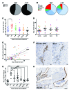
Figure 1. Postpartum breast cancers exhibit increased lymph node metastasis and lymphatic vessel density.
(A) Percentage of lymph node (LN) positivity is increased in women diagnosed with breast cancer within 2 years postpartum (<2 PP) (n = 38) compared with women who have never been pregnant (NBP) (n = 190). (B) Similar percentages of tumor subtypes are observed in postpartum and nulliparous groups: luminal A (LumA), luminal B (LumB), unknown luminal [Lum(unk)] (ER status was positive but PR unknown), Her2, or triple-negative (TN) subtype. (C) Peritumor D2-40+ vessel density is increased in breast cancers diagnosed within 3 years of last childbirth (<3) in comparison to that in women who have never been pregnant (exact P = 0.0186). (D) Increased peritumor D2-40+ vessel density observed in the <3 group is not significantly increased by any known tumor biologic subtype. Color coding shows the reproductive-based tumor categories when separated by subtype. (E) D2-40+ vessel density correlates with number of D2-40+ vessels containing tumor cell nuclei (Pearson r = 0.5811). (F) Representative image of tumor cell lymphatic invasion in a D2-40+/Ki67-stained breast cancer specimen. Tissue sections were stained for Ki67 (right) then for D2-40 (left). (G) D2-40+ lymphatic vessel density is increased in normal adjacent breast tissue from women biopsied less than or equal to 1-year postpartum (≤1), compared with women biopsied 1–3 (>1≤3), 3–6 (>3≤6), 6–10 (>6≤10), 10–15 (>10≤15), and greater than 15 (>15) years postpartum and women who have never been pregnant. (H) Representative image depicting D2-40+ vessels containing cellular debris (arrow) or cells (arrowhead) in breast tissue obtained during 2 weeks after lactation. All data points are depicted along with group average (black bar ± SEM). Scale bar: 10 μm. *P < 0.05, **P < 0.01, ***P < 0.001, ****P < 0.0001, t test.

Comment in

References

    1. Lyons TR, et al. Postpartum mammary gland involution drives progression of ductal carcinoma in situ through collagen and COX-2. Nat Med. 2011;17(9):1109–1115. doi: 10.1038/nm.2416. - DOI - PMC - PubMed
    1. Lyons TR, Schedin PJ, Borges VF. Pregnancy and breast cancer: when they collide. J Mammary Gland Biol Neoplasia. 2009;14(2):87–98. doi: 10.1007/s10911-009-9119-7. - DOI - PMC - PubMed
    1. McDaniel SM, et al. Remodeling of the mammary microenvironment after lactation promotes breast tumor cell metastasis. Am J Pathol. 2006;168(2):608–620. doi: 10.2353/ajpath.2006.050677. - DOI - PMC - PubMed
    1. O’Brien J, et al. Alternatively activated macrophages and collagen remodeling characterize the postpartum involuting mammary gland across species. Am J Pathol. 2010;176(3):1241–1255. doi: 10.2353/ajpath.2010.090735. - DOI - PMC - PubMed
    1. Jindal S, et al. Postpartum breast involution reveals regression of secretory lobules mediated by tissue-remodeling. Breast Cancer Res. 2014;16(2):R31. - PMC - PubMed

Publication types