. 2014 Sep;124(9):3923-8.
doi: 10.1172/JCI75746. Epub 2014 Aug 18.

A human immunodeficiency caused by mutations in the PIK3R1 gene

A human immunodeficiency caused by mutations in the PIK3R1 gene

Marie-Céline Deau et al. J Clin Invest. 2014 Sep.

Erratum in

  • A human immunodeficiency caused by mutations in the PIK3R1 gene.
    Deau MC, Heurtier L, Frange P, Suarez F, Bole-Feysot C, Nitschke P, Cavazzana M, Picard C, Durandy A, Fischer A, Kracker S. Deau MC, et al. J Clin Invest. 2015 Apr;125(4):1764-5. doi: 10.1172/JCI81746. Epub 2015 Apr 1. J Clin Invest. 2015. PMID: 25831445 Free PMC article. No abstract available.

Abstract

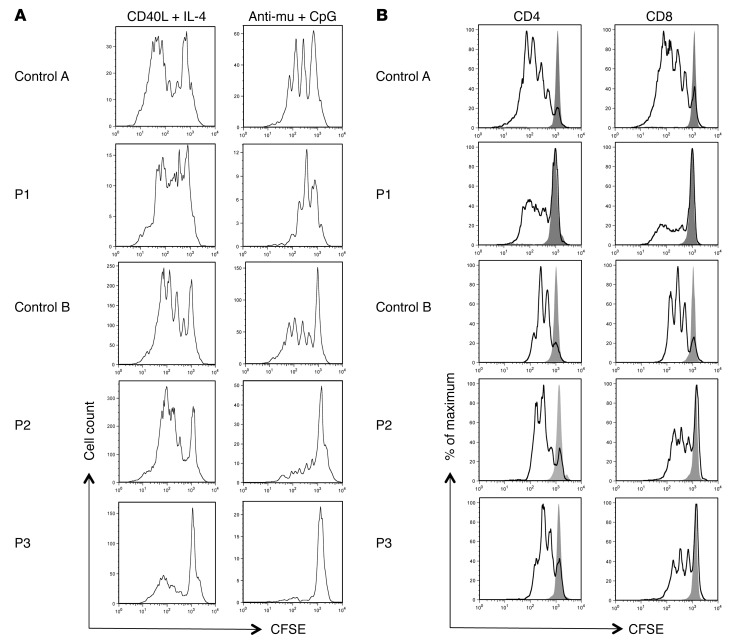
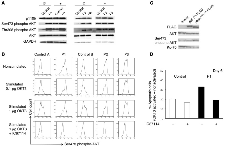
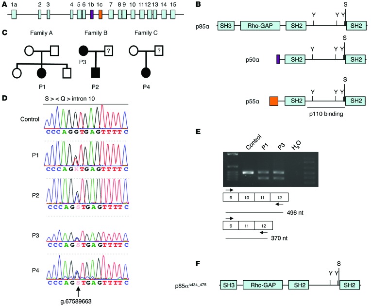
Recently, patient mutations that activate PI3K signaling have been linked to a primary antibody deficiency. Here, we used whole-exome sequencing and characterized the molecular defects in 4 patients from 3 unrelated families diagnosed with hypogammaglobulinemia and recurrent infections. We identified 2 different heterozygous splice site mutations that affect the same splice site in PIK3R1, which encodes the p85α subunit of PI3K. The resulting deletion of exon 10 produced a shortened p85α protein that lacks part of the PI3K p110-binding domain. The hypothetical loss of p85α-mediated inhibition of p110 activity was supported by elevated phosphorylation of the known downstream signaling kinase AKT in patient T cell blasts. Analysis of patient blood revealed that naive T and memory B cell counts were low, and T cell blasts displayed enhanced activation-induced cell death, which was corrected by addition of the PI3Kδ inhibitor IC87114. Furthermore, B lymphocytes proliferated weakly in response to activation via the B cell receptor and TLR9, indicating a B cell defect. The phenotype exhibited by patients carrying the PIK3R1 splice site mutation is similar to that of patients carrying gain-of-function mutations in PIK3CD. Our results suggest that PI3K activity is tightly regulated in T and B lymphocytes and that various defects in the PI3K-triggered pathway can cause primary immunodeficiencies.


Figures

Figure 3
Figure 3. Proliferative ability of peripheral blood B and T lymphocytes.
Proliferation profile as determined by dilution of CFSE-labeled (A) CD19+ gated peripheral blood lymphocytes from patients and the control 5 days after the indicated activation and (B) CD4+ or CD8+ gated peripheral blood lymphocytes from the patients and the control 3 days after stimulation with anti-CD3– and anti-CD28–coated beads. The gray peak indicates CFSE labeling of nonstimulated cells.
Figure 2
Figure 2. Abundance of p110δ protein, phosphorylation of AKT at Ser473, and increased AICD in T cell blasts from patients.
(A) Expression of p110δ, AKT, and GAPDH and phosphorylation of AKT at Ser473 and at Thr308 by Western blotting of cell lysates derived from T cell blasts from P1, P2, and P3 and controls before (Ø) and after (+) stimulation with anti-CD3 antibody. (B) Intracellular staining of phosphorylated AKT at Ser473 in nonstimulated and CD3-activated T cell blasts from patients and controls with or without 10 μM IC87114 inhibitor, measured by flow cytometry. (C) A representative experiment showing the expression of flag-tagged WT p85α, flag-tagged p85αΔ434_475, AKT, and Ku-70 and phosphorylation of AKT at Ser473 by Western blotting of cell lysates derived from NIH3T3 cells 2 days after transfection with the indicated expression vector. (D) Representative AICD experiment with T cell blasts from P1 and the control. The results are expressed as the percentage of apoptotic/dead blasts after OKT3 activation minus the percentage of apoptotic/dead blasts in the absence of stimulation. AICD was analyzed with or without 3 μM IC87114 inhibitor with T cell blasts cultured for 6 days. Similar results were obtained at later time points and for P2 and P3 (data not shown).
Figure 1
Figure 1. A splice site mutation in PIK3R1 leads to deletion of part of the p110-binding domain.
(A) Exons of the PIK3R1 gene, which encodes p85α and the isoforms p50α (purple) and p55α (orange). (B) Protein structure of p85α and the isoforms p50 and p55α. Phosphorylated tyrosine (Y) and serine (S) residues are indicated. (C) Pedigrees of the families carrying PIK3R1 splice site mutations. (D) A sequencing chromatogram showing the heterozygous splice site mutations. Corresponding protein sequence is shown on the top. (E) RT-PCR covering exon 10 of PIK3R1 from indicated samples. (F) Protein structure of p85αΔ434_475 mutant protein.

Comment in

References

    1. Angulo I, et al. Phosphoinositide 3-kinase delta gene mutation predisposes to respiratory infection and airway damage. Science. 2013;342(6160):866–871. doi: 10.1126/science.1243292. - DOI - PMC - PubMed
    1. Lucas CL, et al. Dominant-activating germline mutations in the gene encoding the PI(3)K catalytic subunit p110δ result in T cell senescence and human immunodeficiency. Nat Immunol. 2014;15(1):88–97. - PMC - PubMed
    1. Okkenhaug K, Vanhaesebroeck B. PI3K in lymphocyte development, differentiation and activation. Nat Rev Immunol. 2003;3(4):317–330. doi: 10.1038/nri1056. - DOI - PubMed
    1. Deane JA, Fruman DA. Phosphoinositide 3-kinase: diverse roles in immune cell activation. Annu Rev Immunol. 2004;22:563–598. doi: 10.1146/annurev.immunol.22.012703.104721. - DOI - PubMed
    1. Srinivasan L, et al. PI3 kinase signals BCR-dependent mature B cell survival. Cell. 2009;139(3):573–586. doi: 10.1016/j.cell.2009.08.041. - DOI - PMC - PubMed

Publication types

Substances