. 2014 Sep;124(9):3929-44.
doi: 10.1172/JCI75943. Epub 2014 Aug 18.

PAX7 expression defines germline stem cells in the adult testis

PAX7 expression defines germline stem cells in the adult testis

Gina M Aloisio et al. J Clin Invest. 2014 Sep.

Abstract

Spermatogenesis is a complex, multistep process that maintains male fertility and is sustained by rare germline stem cells. Spermatogenic progression begins with spermatogonia, populations of which express distinct markers. The identity of the spermatogonial stem cell population in the undisturbed testis is controversial due to a lack of reliable and specific markers. Here we identified the transcription factor PAX7 as a specific marker of a rare subpopulation of A(single) spermatogonia in mice. PAX7+ cells were present in the testis at birth. Compared with the adult testis, PAX7+ cells constituted a much higher percentage of neonatal germ cells. Lineage tracing in healthy adult mice revealed that PAX7+ spermatogonia self-maintained and produced expanding clones that gave rise to mature spermatozoa. Interestingly, in mice subjected to chemotherapy and radiotherapy, both of which damage the vast majority of germ cells and can result in sterility, PAX7+ spermatogonia selectively survived, and their subsequent expansion contributed to the recovery of spermatogenesis. Finally, PAX7+ spermatogonia were present in the testes of a diverse set of mammals. Our data indicate that the PAX7+ subset of A(single) spermatogonia functions as robust testis stem cells that maintain fertility in normal spermatogenesis in healthy mice and mediate recovery after severe germline injury, such as occurs after cancer therapy.


Figures
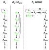
Figure 12
Figure 12. Models of stemness in mouse spermatogenesis.
Spermatogonial subsets proposed as the bona fide stem cells are shown above each of the 3 models. In the classic Asingle model (As), Asingle spermatogonia are homogeneous and share stem cell identity (green), having the capacity for self-maintenance (circular arrows; refs. 3, 4, 66). More recently, models have been proposed arguing for greater plasticity among undifferentiated (Asingle→Aal16) spermatogonia, with chain fragmentation representing one possible mechanism by which stemness is maintained or regenerated (5). Although fragmentation has been shown to occur in vivo, its contributions to stem cell maintenance under normal conditions or after chemotherapy/radiation have not been formally established. Our findings that only a subset of Asingle spermatogonia expressed PAX7 and that these spermatogonia functioned as stem cells suggests a new Asingle subset model, whereby PAX7+ spermatogonia are self-maintaining and may sit atop the hierarchy of spermatogenic differentiation. That Asingle spermatogonia were heterogeneous and that only a subset functioned as stem cells was also suggested by previous studies (10, 67). If so, then this would suggest that some subset of Asingle spermatogonia represent transit-amplifying (TA) intermediates. The number of such transit-amplifying steps between PAX7+ Asingle and Apair spermatogonia is unknown. It will be interesting to determine whether ID4 and ERBB3, expressed in Asingle spermatogonia, are expressed in overlapping or nonoverlapping subsets of spermatogonia relative to PAX7 (9, 49, 50). Other models are possible, such as ones combining different aspects of these models (i.e., fragmentation with the presence of PAX7+ spermatogonia, if fragmentation is confirmed as a functionally significant biological process).
Figure 11
Figure 11. PAX7+ spermatogonia are conserved in mammals.
(A) Epitope mapping of anti-PAX7 mouse monoclonal antibody (generated against chicken aa 320–523). Chicken and corresponding mouse polypeptide aa sequences were tiled as sequential 12-mers at 1-aa resolution. (B) PAX7 Western blot (uncropped to show all visible bands in lanes shown). Addition of blocking peptide (22 aa) confirmed that the anti-PAX7 monoclonal antibody bound to the QPQADFSISP epitope. C2C12, skeletal muscle myoblast cell line. Uterus was included as a negative control. Molecular weight markers denote 75, 50, and 37 kDa. (C) IHC of testes from 7 additional mammalian species, including juveniles for 2 species. PAX7+ spermatogonia (arrows) were rare and localized to the basement membrane. Scale bar: 25 μm.
Figure 10
Figure 10. Pax7 cKO in the male germline.
For each analysis, n = 3 animals were evaluated per genotype. (A) Testes from floxed Pax7fl/fl control and Pax7 cKO (VC;Pax7–/fl) males at 6 months of age. No abnormalities or size differences were noted. (B) Testis weight expressed as percent of total body weight (6 months of age). (C) Fertility assays of 6 month-old males. Bars denote means. All floxed control and Pax7 cKO males were fertile and sired litters of normal size. (D) Histological analyses at 6 months of age. No abnormalities in spermatogenesis or testis morphology were noted. (E) IHC analyses of Pax7 cKO males at PD7. PAX7+ spermatogonia were abundant and present in most tubules in wild-type controls (arrow), but absent in Pax7 cKO tubules (multiple sections were stained and examined for each), confirming the specificity of the PAX7 antibody in testis sections. GCNA shows the presence of germ cells. Scale bars: 50 μm.
Figure 9
Figure 9. Lineage tracing of PAX7+ spermatogonia following busulfan (20 mg/kg) treatment of Pax7-CreERT2;mT/mG males at 6 weeks of age.
(A) Schematic showing both busulfan lineage-tracing experiments. Testes were harvested 8 weeks after the last drug dose for each experiment. (B) Number of clones 8 weeks after busulfan administration. Each point represents 1 testis from 1 animal; red bars denote means; P values were determined by unpaired t test. (C) Clone size 8 weeks after tamoxifen administration. Red bars denote means; P values were determined by unpaired t test. (D) Composite image of representative large clone from tam→bu experiment. Tubule borders are highlighted with dashed lines. Sp, elongate spermatids (arrows denote individual cells or small groupings forming a “trail” of cells). (E) Cryosection of testis from tam→bu experiment showing germ cell clone spanning the entire tubule. ST, seminiferous tubule; LC, Leydig cells. Scale bar: 200 μm (D); 25 μm (E).
Figure 8
Figure 8. Counts of FOXO1+ spermatogonia after busulfan administration at 6 weeks of age.
For each time point, n = 3 animals were analyzed; error bars denote SEM. (A) FOXO1+ spermatogonia per tubule. (B) FOXO1+ counts per tubule compared with PAX7+ counts. (C) IHC showing FOXO1+ spermatogonia (arrows). Unlike PAX7+ spermatogonia, FOXO1+ (undifferentiated) spermatogonia were not resistant to busulfan. Scale bar: 25 μm.
Figure 7
Figure 7. Germline ablation with busulfan has dose-dependent effects on PAX7+ spermatogonia.
(A) Number of cells expressing GCNA (pan-germ cell marker) and PAX7. Error bars denote SEM for n = 3 animals at 6 weeks of age. (B) H&E and immunostained sections 16 days after a single dose of 40 mg/kg busulfan. GCNA stains all germ cells to the round spermatid stage. Busulfan resulted in expansion of PAX7+ clusters never observed in untreated testes; an example of a 4-cell group is shown (insets; enlarged ×4). (C) Fractions of PAX7+ clusters of different sizes. For each time point, fractions add up to 1. The difference in cluster sizes (1 versus ≥2) was highly statistically significant in untreated animals versus those treated with 40 mg/kg busulfan after 32 days (P = 2 × 10⁻⁹). (D) Percent EdU incorporation in PAX7+ spermatogonia 4 days after busulfan administration. Scale bar: 10 μm.
Figure 6
Figure 6. PAX7+ spermatogonia have long-term stem potential in vivo, and their descendants function as stem cells in transplantation assays.
For PD3 time points, tamoxifen injections were performed at PD1 and PD2; for later time points, tamoxifen administration was performed for 3 consecutive days starting at PD3. (A) Clone size. Each column represents 1 testis from separate animals (total n = 15); red bars denote means. Note that many labeled Asingle spermatogonia were present at PD21, demonstrating that PD21 PAX7+ spermatogonia are derived from neonatal PAX7+ spermatogonia. Clones grew over time and persisted in aged animals (12 weeks). (B) Representative clone morphologies by confocal microscopy (n denotes number of cells in labeled chain shown); z stacks confirmed cell counts and Asingle status. (C) Mitotic and apoptotic indices of PAX7+ cells at PD3 (n = 3 animals) demonstrated that early PAX7+ cells were highly proliferative and not characterized by significant apoptosis. Error bars denote SEM. (D) Transplantation assay. A Pax7-CreERT2;tdTomato donor was treated with tamoxifen at PD3. Testes were disaggregated at PD14 and transplanted into germ cell–deficient KitW/KitW–v hosts, which were sacrificed after 4 weeks (n = 3). All hosts (but no controls) showed multiple labeled clones (i.e., 15–20); representative examples are shown. Scale bars: 25 μm (B); 100 μm (D).
Figure 5
Figure 5. Lineage tracing of PAX7+ descendants in Pax7-CreERT2;mT/mG testes.
Adult males were treated with tamoxifen at 6 weeks of age, then aged for the indicated intervals. (A) Clone morphology by confocal microscopy of isolated tubules. Representative Asingle, Apair, Aal4, and Aal8 clones 1 week after tamoxifen administration are shown. Other panels show larger clones at 6 weeks; arrows indicate detached Asingle spermatogonia that were part of larger clones. Inset: elongated spermatid tails in tubular lumen. Clones >500 cells could not be reliably counted. Larger clones were associated with smaller separate chains at their periphery, including Asingle spermatogonia. The few 1-cell clones at 6–16 weeks represent Asingle spermatogonia too distant from the nearest clone to be confidently identified as part of it, but may reflect long-distance migration. Green motile sperm were observed in the epididymis. (B) Clone morphology in tissue sections (16 weeks after tamoxifen). PAX7+ descendants gave rise to all spermatogenic stages, as evidenced by circumferential full-thickness labeling of all spermatogenic stages throughout the tubule. Tissue sections were counterstained with DAPI. (C) Labeled sperm from epididymis showing bright green fluorescence and characteristic hook morphology. The majority of sperm did not exhibit fluorescence, and control epididymides did not contain spermatozoa with comparable fluorescence (i.e., the signal shown is not background autofluorescence of sperm). (D) Average clone number in n = 4 testes. Clone numbers did not decrease over time. (E) Clone size. Red bars denote means. Larger clones included detached smaller chains and Asingle spermatogonia. Scale bars: 25 μm.
Figure 4
Figure 4. Lineage tracing of PAX7+ descendants in Pax7-CreERT2;R26R testes.
Mice were treated with tamoxifen at 6 weeks of age then aged for the indicated intervals. (A) Representative clones at 4 days, 3 weeks, and 6 weeks. Dashed lines demarcate tubule borders. Arrows denote a 1-cell clone at 4 days or isolated Asingle spermatogonia at the periphery of larger clones at 6 weeks. All marker-expressing cells in these fields are considered clonal because there were no additional marked cells in the tubule. The H&E-stained section of Xgal-treated testis at 6 weeks showed a single-labeled (blue) elongate spermatid tail among many unlabeled (pink) spermatid tails in the tubular lumen. Other labeled spermatid tails were outside the field of view. Scale bars: 100 μm; 10 μm (H&E). (B) Average clone numbers in n = 4 testes. The 1-week time point may represent undercount due to difficulty in visualizing single-cell clones, or marker expression lag. Clone numbers were consistent with the presence of approximately 400 PAX7+ cells per testis and 10% recombination efficiency for Pax7-CreERT2 after tamoxifen administration (31). (C) Clone size. Red bars denote means. Larger clones were composed of large labeled zones and peripheral smaller chains including Asingle spermatogonia.
Figure 3
Figure 3. PAX7+ spermatogonia make up a higher percentage of germ cells in the neonatal testis.
(A) PAX7+ cell frequency, expressed as a percentage of total GCNA+ cells. Inset: PAX7 IHC at PD7, showing several PAX7+ spermatogonia (asterisks). Magnification, ×250. (B) Counts of total PAX7+ cells by IHC of serially sectioned PD1 and adult testes (n = 3). Each bar represents a single testis. Total numbers were similar in neonatal and adult testes. (C) 6-week-old animals were injected with EdU, and testes were harvested after the indicated times. The experiment was repeated twice with similar results.
Figure 2
Figure 2. PAX7+ spermatogonia in normal testes.
(A) Rarity of PAX7+ cells in adult (6-week-old) testis cross-section. Arrow denotes a PAX7+ cell within a single seminiferous tubule. (B) Relative number of spermatogonia positive for known markers compared with PAX7 in tissue sections, with >90 tubules counted per testis. Error bars denote SEM of averages derived from 3 6-week-old animals. (C) Double-labeling (confocal microscopy) showing that PAX7+ cells were FOXO1+ and GFRα1+ (representative examples among ≥10 PAX7+ cells). Basement membrane staining is nonspecific. (D) FOXO1 and PAX7 double-labeling of Asingle, Apair, and Aal4–Aal16 chains, visualized by confocal microscopy. PAX7+ spermatogonia were Asingle spermatogonia; larger chains did not contain PAX7+ spermatogonia. Arrow indicates an Aal4 chain. Bottom panels show different channels for the same field of a single PAX7+FOXO1+ spermatogonium (asterisk in DAPI channel). (E) KIT and PAX7 double-labeling (confocal microscopy) showed that PAX7+ spermatogonia (arrow) were isolated (i.e., Asingle) and KIT. No KIT+PAX7+ spermatogonia were observed. PAX7 was nuclear, whereas KIT was membrane-associated, as expected. Image shows 3 tubules optically sectioned close to the level of the basement membrane to visualize large numbers of spermatogonia. Scale bars: 10 μm (A); 25 μm (C and D); 50 μm (E).
Figure 1
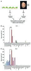
Figure 1. Digital Northern analysis identifying PAX7 as potential adult testis germline stem cell marker.
(A) General RNA-based approach to identify markers that were highly expressed in cultured SSCs relative to adult testis. (B and C) Relative expression levels of (B) Pax7 and (C) Ddx4 across multiple samples. Error bars denote SEM. Pax7 mRNA levels were >180-fold higher in established spermatogonial cultures relative to adult testis. SSC, cultured SSCs; Sl/Sl(d), KitlSl/KitlSl–d germ cell–deficient adult testes; ES, embryonic stem; HS, hematopoietic stem.

Comment in

References

    1. Oatley JM, Brinster RL. Regulation of spermatogonial stem cell self-renewal in mammals. Annu Rev Cell Dev Biol. 2008;24:263–286. doi: 10.1146/annurev.cellbio.24.110707.175355. - DOI - PMC - PubMed
    1. de Rooij DG. Proliferation and differentiation of spermatogonial stem cells. Reproduction. 2001;121(3):347–354. doi: 10.1530/rep.0.1210347. - DOI - PubMed
    1. de Rooij DG, Russell LD. All you wanted to know about spermatogonia but were afraid to ask. J Androl. 2000;21(6):776–798. - PubMed
    1. Oakberg EF. Spermatogonial stem-cell renewal in the mouse. Anat Rec. 1971;169(3):515–531. doi: 10.1002/ar.1091690305. - DOI - PubMed
    1. Nakagawa T, Sharma M, Nabeshima Y, Braun RE, Yoshida S. Functional hierarchy and reversibility within the murine spermatogenic stem cell compartment. Science. 2010;328(5974):62–67. doi: 10.1126/science.1182868. - DOI - PMC - PubMed

Publication types

Substances