. 2014 Sep;124(9):4102-14.
doi: 10.1172/JCI76069. Epub 2014 Aug 18.

A selective microRNA-based strategy inhibits restenosis while preserving endothelial function

A selective microRNA-based strategy inhibits restenosis while preserving endothelial function

Gaetano Santulli et al. J Clin Invest. 2014 Sep.

Abstract

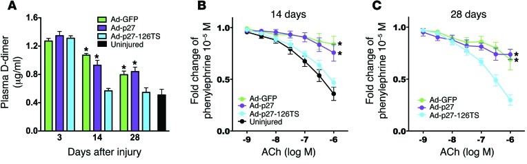
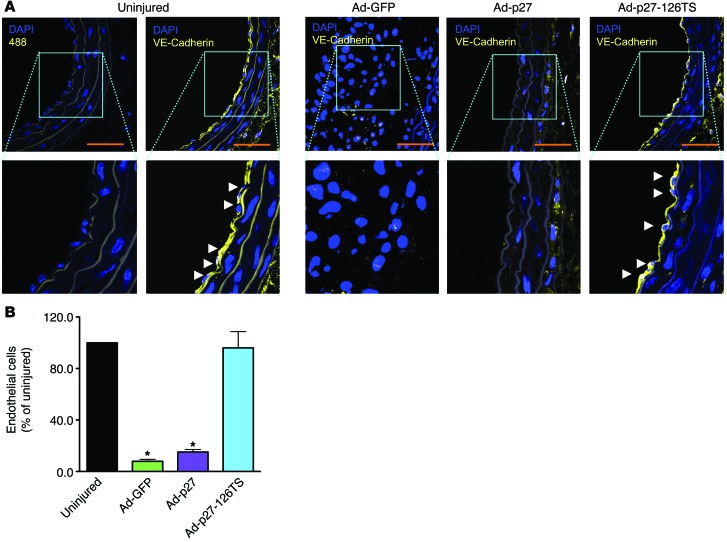
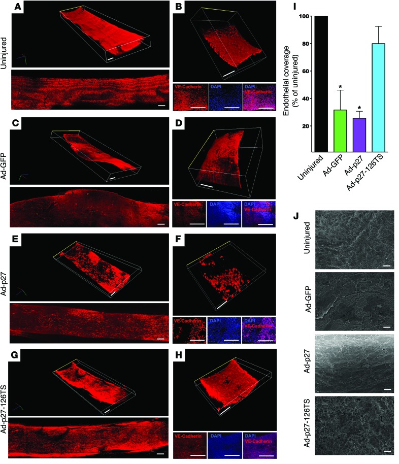
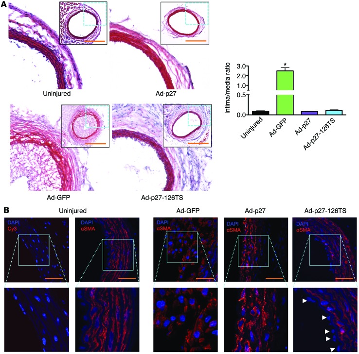
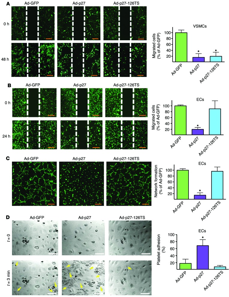
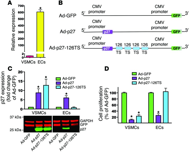
Drugs currently approved to coat stents used in percutaneous coronary interventions do not discriminate between proliferating vascular smooth muscle cells (VSMCs) and endothelial cells (ECs). This lack of discrimination delays reendothelialization and vascular healing, increasing the risk of late thrombosis following angioplasty. We developed a microRNA-based (miRNA-based) approach to inhibit proliferative VSMCs, thus preventing restenosis, while selectively promoting reendothelialization and preserving EC function. We used an adenoviral (Ad) vector that encodes cyclin-dependent kinase inhibitor p27(Kip1) (p27) with target sequences for EC-specific miR-126-3p at the 3' end (Ad-p27-126TS). Exogenous p27 overexpression was evaluated in vitro and in a rat arterial balloon injury model following transduction with Ad-p27-126TS, Ad-p27 (without miR-126 target sequences), or Ad-GFP (control). In vitro, Ad-p27-126TS protected the ability of ECs to proliferate, migrate, and form networks. At 2 and 4 weeks after injury, Ad-p27-126TS-treated animals exhibited reduced restenosis, complete reendothelialization, reduced hypercoagulability, and restoration of the vasodilatory response to acetylcholine to levels comparable to those in uninjured vessels. By incorporating miR-126-3p target sequences to leverage endogenous EC-specific miR-126, we overexpressed exogenous p27 in VSMCs, while selectively inhibiting p27 overexpression in ECs. Our proof-of-principle study demonstrates the potential of using a miRNA-based strategy as a therapeutic approach to specifically inhibit vascular restenosis while preserving EC function.


Figures

Figure 6
Figure 6. Ad-p27-126TS reduces hypercoagulability and restores EC function in vivo.
(A) Plasma D-dimer levels evaluated before (uninjured) and 3, 14, and 28 days after balloon injury using a specific immunoassay. n = 5 animals/group. (B and C) Vascular reactivity analysis on carotid rings 2 (B) and 4 (C) weeks after injury showing the vasodilatory response to acetylcholine (ACh). n = 5–6 rats/group. Data represent the means ± SEM and were compared using 1-way (A) or 2-way repeated measures (B and C) ANOVA followed by Tukey-Kramer’s post hoc test. *P < 0.01 versus uninjured arteries.
Figure 5
Figure 5. Ad-p27-126TS restores reendothelialization in cross sections of carotid arteries.
(A) Representative sections of rat carotid arteries immunostained for the specific EC marker VE-cadherin 2 weeks after injury. Nuclei were counterstained with DAPI. No positive staining was observed in the negative control sections (Alexa Fluor 488 alone). Scale bars: 100 μm; original magnification, ×60 and ×120 (insets). Arrowheads indicate ECs beyond the inner autofluorescent elastic laminae. (B) Endothelial coverage was quantified by counting the number of VE-cadherin–positive cells in the circumference of the lumens from at least 6 animals/group. Data were compared using 1-way ANOVA with Tukey-Kramer’s post hoc test. *P < 0.05 versus uninjured arteries.
Figure 4
Figure 4. Ad-p27-126TS restores endothelial coverage of en face longitudinal arterial preparations.
(AH) Representative confocal images of the internal surface of the vessels immunostained for VE-cadherin. (A, C, E, and G) Tridimensional (top) and bidimensional (bottom) images of the longitudinal en face preparation of the carotid arteries. (B, D, F, and H) Tridimensional images of a representative central portion (see Methods and Supplemental Figure 7A for detailed information about the imaging procedure) of the vessel (top), with the respective 2D pictures showing the EC-specific immunostaining for VE-cadherin (bottom: VE-cadherin, DAPI, and merge) and representing the endothelial coverage, which is quantified in I; the luminal side is indicated by a yellow line. See also the Supplemental Videos 1–8 for a tridimensional view. Dimensional scale bars: 400 μm (all 2D and 3D images). Data comparisons were made using 1-way ANOVA with Tukey-Kramer’s post hoc test; n = at least 5/group. *P < 0.01 versus uninjured arteries. (J) Representative scanning electron microscopy images of the internal surface of uninjured, Ad-GFP–, Ad-p27–, and Ad-p27-126TS–transduced arteries. Scale bars: 10 μm.
Figure 3
Figure 3. Ad-p27 and Ad-p27-126TS inhibit restenosis 2 weeks after balloon injury.
(A) Representative H&E-stained sections. Scale bars: 500 μm; original magnification, ×10 (insets show the whole arterial section at ×5 original magnification). Intima/media ratios were calculated from at least 6 rats/group. Data represent the means ± SEM. Data comparisons were made using 1-way ANOVA with Tukey-Kramer’s post hoc test. *P < 0.01 versus uninjured arteries. (B) Representative sections of rat carotid arteries immunostained for α-SMA. Nuclei were counterstained with DAPI. No positive staining was observed in the negative control sections. Scale bars: 100 μm; original magnification, ×60 and ×120 (insets). Arrowheads indicate EC nuclei beyond the inner autofluorescent elastic laminae.
Figure 2
Figure 2. Ad-p27-126TS inhibits VSMC migration while preserving EC properties in vitro.
Migration assays of VSMCs (A) and ECs (B) transduced with the indicated Ad vector. Representative images at the indicated time points are shown. Wound areas were measured using ImageJ64 software. (C) Formation of network-like structures in ECs was quantified by counting branch points. Scale bars: 100 μm (AC). Data represent the means ± SEM of at least 3 experiments performed in quadruplicate. (D) Representative photomicrographs of platelet adhesion to ECs transduced with the indicated Ad vector in a parallel-plate flow chamber apparatus before (upper panels, t = 0) and after perfusion of PPACK-treated blood and washing (lower panels, t = 3 minutes). Yellow arrows represent human platelets. Scale bars: 30 μm. Graph on the right represents a quantification of the platelet adhesion test performed using Fiji software. Data represent the means ± SEM of at least 3 experiments performed in triplicate. Data comparisons were made using 1-way ANOVA with Tukey-Kramer’s post hoc test. *P < 0.01 versus Ad-GFP.
Figure 1
Figure 1. miR-126-3p target sequences selectively protect ECs from the antiproliferative effects of p27.
(A) RT-qPCR analysis of miR-126-3p expression. (B) Schematic representation of the p27 expression cassette in the Ad vectors engineered to inhibit overexpression of exogenous p27 in cells expressing miR-126-3p (i.e., ECs). (C) Representative immunoblot of p27 protein expression from 3 independent experiments accompanied by densitometric quantification. Lanes were run on the same gel but were noncontiguous. For each cell type, p27 expression was normalized to each vector’s GFP expression to account for transduction efficiency. (D) Proliferation assays of VSMCs and ECs transduced with the indicated Ad vector. Data represent the mean ± SEM of at least 3 experiments performed in quadruplicate. Data comparisons were made using 1-way ANOVA with Tukey-Kramer’s post hoc test. *P < 0.01 versus VSMCs (A); *P < 0.01 versus Ad-GFP (C and D).

Comment in

References

    1. Stefanini GG, Holmes DR, Holmes DR., Jr Drug-eluting coronary-artery stents. N Engl J Med. 2013;368(3):254–265. doi: 10.1056/NEJMra1210816. - DOI - PubMed
    1. Nabel EG, Braunwald E. A tale of coronary artery disease and myocardial infarction. N Engl J Med. 2012;366(1):54–63. doi: 10.1056/NEJMra1112570. - DOI - PubMed
    1. De Luca G, et al. Drug-eluting vs bare-metal stents in primary angioplasty: a pooled patient-level meta-analysis of randomized trials. Arch Intern Med. 2012;172(8):611–621. doi: 10.1001/archinternmed.2012.758. - DOI - PubMed
    1. Chaabane C, Otsuka F, Virmani R, Bochaton-Piallat ML. Biological responses in stented arteries. Cardiovasc Res. 2013;99(2):353–363. doi: 10.1093/cvr/cvt115. - DOI - PubMed
    1. Marx SO, Totary-Jain H, Marks AR. Vascular smooth muscle cell proliferation in restenosis. Circ Cardiovasc Interv. 2011;4(1):104–111. doi: 10.1161/CIRCINTERVENTIONS.110.957332. - DOI - PMC - PubMed

Publication types

Substances