

Review
doi: 10.1021/bc500320j.
Epub 2014 Sep 2.
Promises and pitfalls of intracellular delivery of proteins
Affiliations
- PMID: 25133522
- PMCID: PMC4166028
- DOI: 10.1021/bc500320j
Item in Clipboard
Review
Promises and pitfalls of intracellular delivery of proteins
Bioconjug Chem.
.
Abstract
The direct delivery of functional proteins into the cell cytosol is a key issue for protein therapy, with many current strategies resulting in endosomal entrapment. Protein delivery to the cytosol is challenging due to the high molecular weight and the polarity of therapeutic proteins. Here we review strategies for the delivery of proteins into cells, including cell-penetrating peptides, virus-like particles, supercharged proteins, nanocarriers, polymers, and nanoparticle-stabilized nanocapsules. The advantages and disadvantages of these approaches including cytosolar delivery are compared and contrasted, with promising pathways forward identified.
Figures
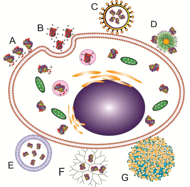
Schematic illustration
of protein delivery systems. (A) cell-penetrating
peptides, (B) supercharged proteins, (C) virus-like particle, (D)
nanocarrier, (E) liposomes, (F) polymer, and (G) nanoparticle-stabilized
nanocapsule.
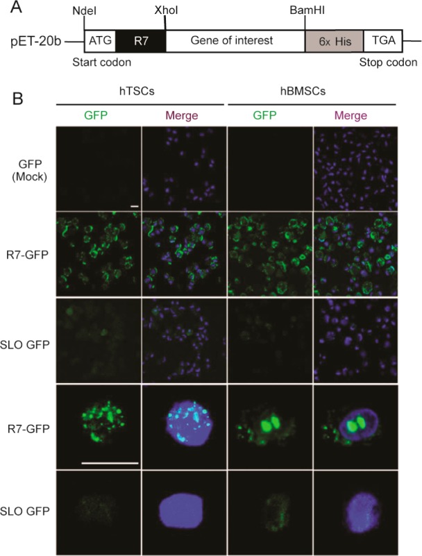
Protein delivery into
cells by using a CPP (R7). (A) Schematic
diagrams of recombinant proteins with or without the CPP (R7)-conjugated
vectors. (B) Comparison of the efficiency of two different protein-delivery
systems (CPP- and Streptolysin O-mediated). Transduction of GFP and
R7-GFP was detected by confocal microscopy. GFP or R7-GFP is visualized
in green. Nuclei were counter-stained with DAPI and the images were
merged (the top three rows show 400× magnification and the bottom
two rows show 1000× magnification plus 3× zoom). Scale bars
represent 20 μm. GFP, green fluorescent protein; DAPI, 4′,6-diamidino-2-phenylindole.
Reprinted with permission from ref (11). Copyright 2011 Nature Publishing Group.
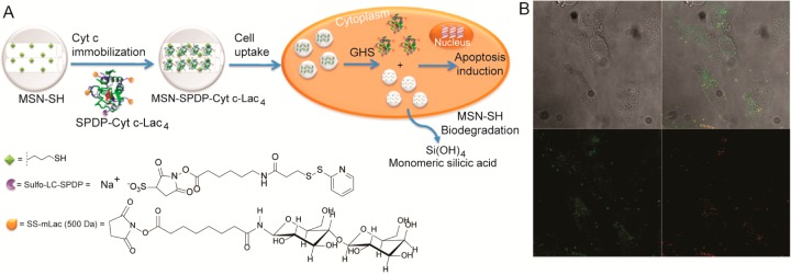
MNPs deliver serratiopeptidase
into cells. (A) Scheme of the immobilization
of Cytc-Lac into MSN-SH via redox-sensitive smart bonds followed by
its intracellular delivery into cancer cells. (B) Internalization
of the MSN-SPDP-Cyt c-Lac bioconjugate by HeLa cells observed by confocal
microscopy. The left image is the autofluorescence image of the cells,
the lower left shows the FITC labeled MSN internalized by the cells,
the lower right shows the FM4-64 labeled endosomes, and the upper
right micrograph is the merged image. FITC, fluorescein isothiocyanate.
Reprinted with permission from ref (36). Copyright 2014 American Chemical Society.
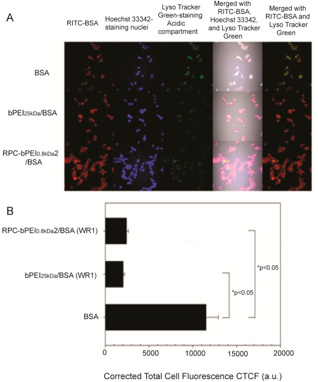
PEI-based
polyelectrolytes deliver protein into MCF7 cells. (A)
Confocal images for intracellular tracking of the cationic polyelectrolyte/BSA
complexes (WR1) and BSA (10 μg/mL) and (B) the intracellular
endolysosome amount of the BSA- and polycation/BSA-treated cells.
Free BSA was labeled with RITC (red). Acidic compartments and nuclei
were stained with LysoTracker Green (green) and Hoechst 33342 (blue),
respectively. RITC, Rhodamine Bisothiocyanate. Reprinted with permission
from ref (51). Copyright
2013 American Chemical Society.
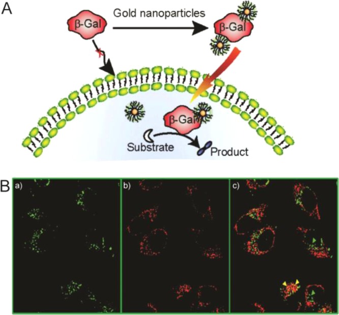
Nanoplex delivery of a large anionic protein
β-galactosidase
(473 kDa) into cells. (A) Intracellular delivery of functional protein
using gold nanoparticles. (B) Co-localization study using confocal
microscopy after protein transfection (NP_Pep/FITC-gal: 100 nM/50
nM) of HeLa cells in the presence of FM4-64: (a) green fluorescence
from FITC-gal, (b) red fluorescence from FM4-64, an endosome-specific
marker, and (c) overlap of the green and the red channels. In the
merged image, green spots (shown with green arrowheads) indicate proteins
outside endosomes, while entrapped proteins inside endosomes appear
as yellow dots (shown with yellow arrowheads). Reprinted with permission
from ref (57). Copyright
2010 American Chemical Society.
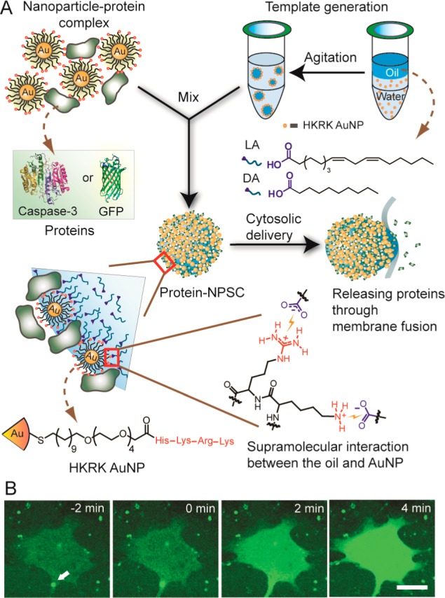
Intracellular
protein delivery by NPSCs. (A) Schematic showing
the preparation of the protein NPSC complex containing caspase-3 or
GFP and proposed delivery mechanism. (B) Live cell imaging of rapid
GFP release into the cytosol of HeLa cell by NPSCs. Scale bar: 20
μm. Reprinted with permission from ref (62). Copyright 2013 American
Chemical Society.
References
-
- Shah D. A.; Kwon S. J.; Bale S. S.; Banerjee A.; Dordick J. S.; Kane R. S. (2011) Regulation of stem cell signaling by nanoparticle-mediated intracellular protein delivery. Biomaterials 32, 3210–3219. - PubMed
-
- Vasconcelos L.; Pärn K.; Langel U. (2013) Therapeutic potential of cell-penetrating peptides. Ther. Delivery 4, 573–591. - PubMed
-
- Kratz F.; Elsadek B. (2012) Clinical impact of serum proteins on drug delivery. J. Controlled Release 161, 429–445. - PubMed
Publication types
MeSH terms
Substances
Grants and funding
LinkOut - more resources
Full Text Sources
Other Literature Sources
