

Impaired cerebrovascular reactivity in multiple sclerosis
- PMID: 25133874
- PMCID: PMC4376108
- DOI: 10.1001/jamaneurol.2014.1668
Impaired cerebrovascular reactivity in multiple sclerosis
Abstract
Importance: Cerebrovascular reactivity (CVR) is an inherent indicator of the dilatory capacity of cerebral arterioles for a vasomotor stimulus for maintaining a spontaneous and instant increase of cerebral blood flow (CBF) in response to neural activation. The integrity of this mechanism is essential to preserving healthy neurovascular coupling; however, to our knowledge, no studies have investigated whether there are CVR abnormalities in multiple sclerosis (MS).
Objective: To use hypercapnic perfusion magnetic resonance imaging to assess CVR impairment in patients with MS.
Design, setting, and participants: A total of 19 healthy volunteers and 19 patients with MS underwent perfusion magnetic resonance imaging based on pseudocontinuous arterial spin labeling to measure CBF at normocapnia (ie, breathing room air) and hypercapnia. The hypercapnia condition is achieved by breathing 5% carbon dioxide gas mixture, which is a potent vasodilator causing an increase of CBF.
Main outcomes and measures: Cerebrovascular reactivity was calculated as the percent increase of normocapnic to hypercapnic CBF normalized by the change in end-tidal carbon dioxide, which was recorded during both conditions. Group analysis was performed for regional and global CVR comparison between patients and controls. Regression analysis was also performed between CVR values, lesion load, and brain atrophy measures in patients with MS.
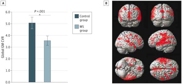
Results: A significant decrease of mean (SD) global gray matter CVR was found in patients with MS (3.56 [0.81]) compared with healthy controls (5.08 [1.56]; P = .001). Voxel-by-voxel analysis showed diffuse reduction of CVR in multiple regions of patients with MS. There was a significant negative correlation between gray matter CVR and lesion volume (R = 0.6, P = .004) and a significant positive correlation between global gray matter CVR and gray matter atrophy index (R = 0.5, P = .03).
Conclusions and relevance: Our quantitative imaging findings suggest impairment in functional cerebrovascular pathophysiology, by measuring a diffuse decrease in CVR, which may be the underlying cause of neurodegeneration in MS.
Figures


Comment in
-
Intracranial relationship between arterioles and venules size.JAMA Neurol. 2015 Jan;72(1):124. doi: 10.1001/jamaneurol.2014.3514. JAMA Neurol. 2015. PMID: 25581862 No abstract available.
-
Intracranial relationship between arterioles and venules size--reply.JAMA Neurol. 2015 Jan;72(1):124-5. doi: 10.1001/jamaneurol.2014.3517. JAMA Neurol. 2015. PMID: 25581863 No abstract available.
References
-
- Brown GC, Borutaite V. Nitric oxide inhibition of mitochondrial respiration and its role in cell death. Free Radic Biol Med. 2002;33(11):1440–1450. - PubMed
-
- Tzourio-Mazoyer N, Landeau B, Papathanassiou D, et al. Automated anatomical labeling of activations in SPM using a macroscopic anatomical parcellation of the MNI MRI single-subject brain. Neuroimage. 2002;15(1):273–289. - PubMed
-
- Zhang Y, Brady M, Smith S. Segmentation of brain MR images through a hidden Markov random field model and the expectation-maximization algorithm. IEEE Trans Med Imaging. 2001;20(1):45–57. - PubMed
-
- Lu H, Golay X, Pekar JJ, Van Zijl PC. Sustained poststimulus elevation in cerebral oxygen utilization after vascular recovery. J Cereb Blood Flow Metab. 2004;24(7):764–770. - PubMed
Publication types
MeSH terms
Substances
Grants and funding
LinkOut - more resources
Full Text Sources
Other Literature Sources
