Controlled Clinical Trial
. 2014 Sep 15;190(6):639-48.
doi: 10.1164/rccm.201403-0505OC.

Epithelial interleukin-25 is a key mediator in Th2-high, corticosteroid-responsive asthma

Affiliations
Controlled Clinical Trial

Epithelial interleukin-25 is a key mediator in Th2-high, corticosteroid-responsive asthma

Dan Cheng et al. Am J Respir Crit Care Med. .

Abstract

Rationale: Activation of type 2 cytokine pathways plays a central role in a large subset of subjects with asthma. Th2-high and Th2-low asthma have distinct clinical, pathologic, and molecular phenotypes and respond differently to therapy. The factors that initiate type 2 responses in some subjects with asthma are unknown.

Objectives: To determine whether expression of epithelial cytokines IL-25, IL-33, and thymic stromal lymphopoietin are associated with type 2 responses and predict response to inhaled corticosteroid (ICS) in asthma.

Methods: We analyzed pulmonary function tests, blood, and bronchoscopic biopsies from 21 healthy control subjects and 43 subjects with asthma. Subjects with asthma underwent an 8-week treatment with inhaled budesonide.

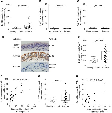
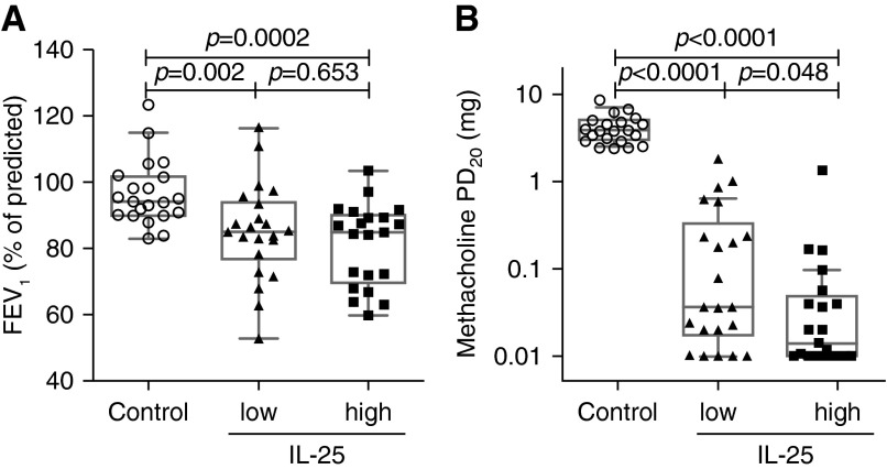
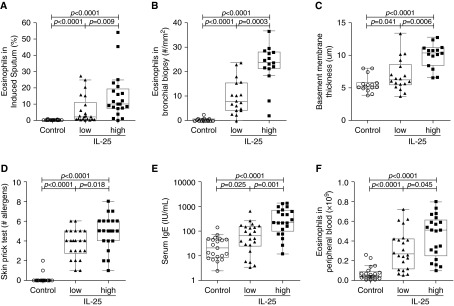
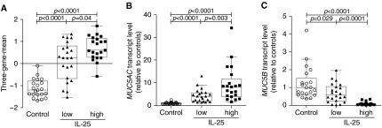
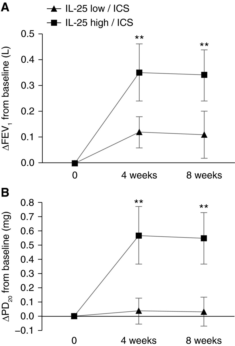
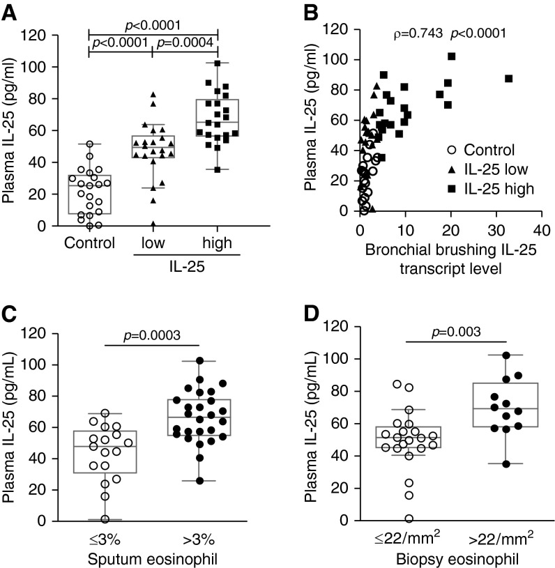
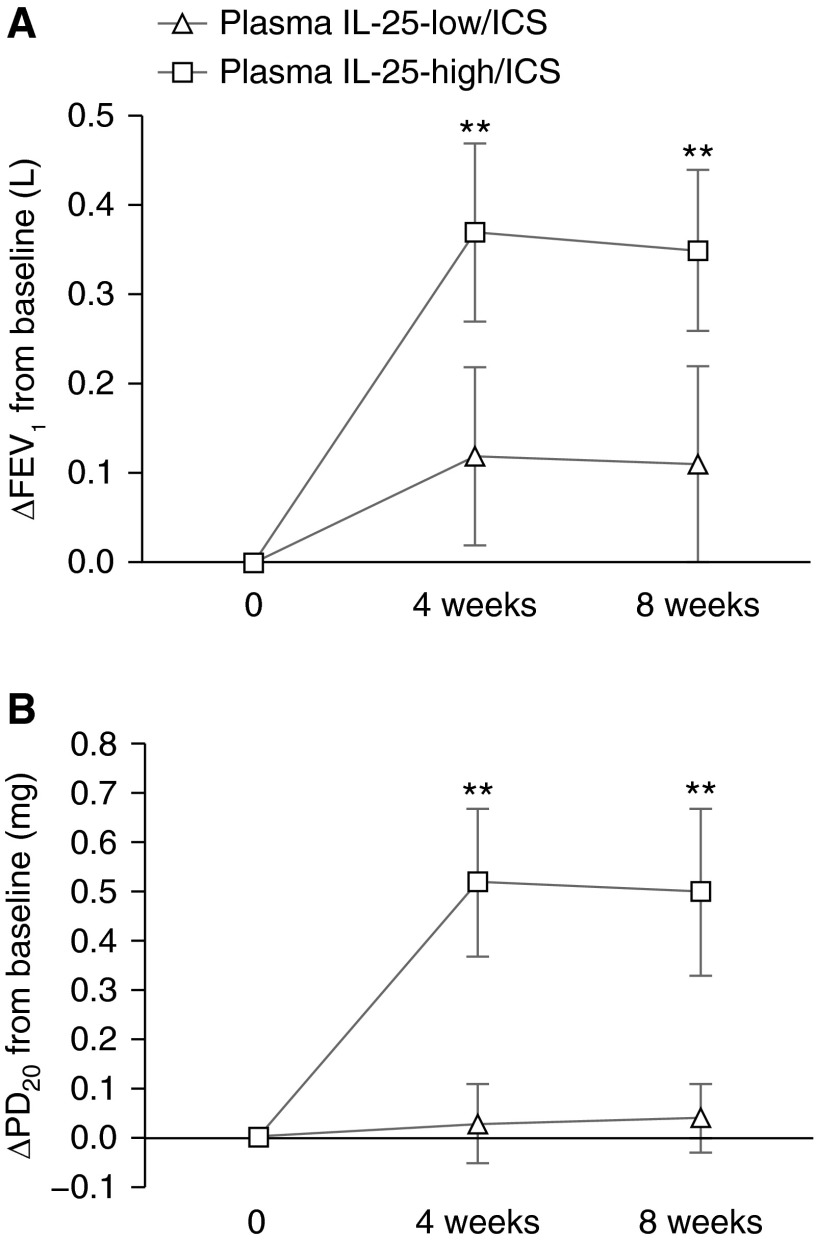
Measurements and main results: Epithelial expression of IL-25, but not IL-33 or thymic stromal lymphopoietin, was increased in a subset of subjects with asthma. The IL-25-high subset had greater airway hyperresponsiveness, more airway and blood eosinophils, higher serum IgE, more subepithelial thickening, and higher expression of Th2 signature genes. ICS improved FEV1 and hyperresponsiveness in the IL-25-high but not the IL-25-low subset. Plasma IL-25 levels correlated with epithelial IL-25 expression, airway eosinophilia, and beneficial responses to ICS treatment.

Conclusions: IL-25 measurements identify two subsets of subjects with distinct asthma phenotypes and different responses to ICS. Because IL-25 has a major role in triggering type 2 responses, bronchial epithelial IL-25 expression is likely a key determinant of type 2 response activation in asthma. Plasma IL-25 level reflects airway IL-25/type 2 response activation and may be useful for predicting responses to asthma therapy.

Keywords: IL-25; asthma; phenotype.


Figures

Figure 1.
Figure 1.
Bronchial epithelial expression of IL-25 and its receptor are increased in some subjects with asthma. Levels of IL-25 (A), IL-33 (B), and thymic stromal lymphopoietin (TSLP) (C) transcripts were determined by quantitative polymerase chain reaction (PCR) of RNA from bronchial brushings of healthy control subjects (n = 21) and subjects with asthma (n = 43). Values are relative to the median value for healthy control subjects. The median IL-25 cycle threshold values for healthy control subjects and asthma subjects were 24.06 and 22.14, respectively. (D) IL-25 immunohistochemistry in bronchial biopsies from healthy control subjects and subjects with asthma (original magnification ×400). (E) Quantitation of IL-25–containing cells in the epithelium of healthy control subjects (n = 13) and subjects with asthma (n = 27). (F) Correlation between bronchial brushing IL-25 transcript level and the number of IL-25–containing cells in airway epithelium for healthy control subjects (n = 13) and subjects with asthma (n = 27). (G) The transcript level of IL-25 receptor, IL-17RB, as determined by quantitative PCR of RNA from bronchial biopsies of healthy control subjects (n = 9) and subjects with asthma (n = 17). (H) Correlation between bronchial brushing IL-25 transcript level and IL-17RB transcript level in bronchial biopsies of healthy control subjects (n = 9) and subjects with asthma (n = 17). Some subjects were not included in the analyses of IL-25 protein and/or IL-17RB transcript level because bronchial biopsies from those subjects were not adequate for reverse transcriptase PCR and/or immunohistochemistry because of limitations in the number of biopsies obtained or the amount of intact tissue seen in the sections.
Figure 2.
Figure 2.
Pulmonary function test results in IL-25-low and IL-25-high asthma. (A) FEV1, a measure of airway obstruction. (B) Methacholine-provocative dosage required to cause a 20% decline in FEV1 (PD20), a measure of airway hyperresponsiveness.
Figure 3.
Figure 3.
Measures of airway eosinophilic inflammation, airway remodeling, and allergy are increased in subjects with IL-25-high asthma. (A) Eosinophils as a percentage of total cells in induced sputum. (B) The number of eosinophils per square millimeter of submucosa in bronchial biopsy. (C) Reticular basement membrane thickness, a measure of subepithelial fibrosis. (D) Skin prick test using a panel of 14 aeroallergens. (E) Serum IgE. (F) Peripheral blood eosinophil count.
Figure 4.
Figure 4.
Bronchial epithelial IL-25 transcript levels are associated with the epithelial expression of Th2 signature genes and mucins. (A) The three-gene-mean derived from the transcript levels of Th2 signature gene CLCA1, POSTN, and SERPINB2 as determined by quantitative polymerase chain reaction in bronchial brushings. (B, C) Levels of transcripts for the major airway mucins MUC5AC (B) and MUC5B (C) in bronchial brushings as determined by quantitative polymerase chain reaction. Values are relative to the median value for healthy control subjects.
Figure 5.
Figure 5.
Subjects with IL-25-high asthma have larger improvements in pulmonary function during inhaled corticosteroid (ICS) treatment. Twenty-two IL-25-low subjects and 21 IL-25-high subjects (as determined by epithelial IL-25 mRNA level) with asthma all received ICS treatment. FEV1 (A) and provocative dosage required to cause a 20% decline in FEV1 (PD20) (B) were measured at baseline (0) and after 4 or 8 weeks on daily inhaled budesonide (200 μg twice a day). **P < 0.01 for IL-25-low/ICS versus IL-25-high/ICS.
Figure 6.
Figure 6.
Increased plasma IL-25 levels are associated with increased bronchial epithelial IL-25 expression and airway eosinophilia. (A) Plasma IL-25 level in peripheral blood as determined by ELISA. (B) Correlation between bronchial brushing IL-25 transcript levels and plasma IL-25 levels of all subjects, including healthy control subjects and subjects with asthma. (C) Plasma IL-25 levels in subjects with asthma with and without sputum eosinophilia. (D) Plasma levels in subjects with asthma and without submucosal eosinophilia in bronchial biopsies.
Figure 7.
Figure 7.
Subjects with elevated plasma IL-25 have larger improvements in pulmonary function during inhaled corticosteroid (ICS) treatment. Twenty plasma IL-25-low subjects (≤55 pg/ml) and 23 plasma IL-25-high subjects (>55 pg/ml) with asthma all received ICS treatment. FEV1 (A) and provocative dosage required to cause a 20% decline in FEV1 (PD20) (B) were measured at baseline (0) and after 4 or 8 weeks on daily inhaled budesonide (200 μg twice a day). **P < 0.01 for plasma IL-25-low/ICS versus plasma IL-25-high/ICS.

Comment in

References

    1. Wenzel SE, Schwartz LB, Langmack EL, Halliday JL, Trudeau JB, Gibbs RL, Chu HW. Evidence that severe asthma can be divided pathologically into two inflammatory subtypes with distinct physiologic and clinical characteristics. Am J Respir Crit Care Med. 1999;160:1001–1008. - PubMed
    1. Green RH, Brightling CE, Woltmann G, Parker D, Wardlaw AJ, Pavord ID. Analysis of induced sputum in adults with asthma: identification of subgroup with isolated sputum neutrophilia and poor response to inhaled corticosteroids. Thorax. 2002;57:875–879. - PMC - PubMed
    1. Woodruff PG, Modrek B, Choy DF, Jia G, Abbas AR, Ellwanger A, Koth LL, Arron JR, Fahy JV. T-helper type 2-driven inflammation defines major subphenotypes of asthma. Am J Respir Crit Care Med. 2009;180:388–395. - PMC - PubMed
    1. Simpson JL, Scott R, Boyle MJ, Gibson PG. Inflammatory subtypes in asthma: assessment and identification using induced sputum. Respirology. 2006;11:54–61. - PubMed
    1. Peters MC, Mekonnen ZK, Yuan S, Bhakta NR, Woodruff PG, Fahy JV.Measures of gene expression in sputum cells can identify Th2-high and Th2-low subtypes of asthma J Allergy Clin Immunol 2013133388–394.e5 - PMC - PubMed

Publication types