. 2014 Nov:76:73-83.
doi: 10.1016/j.yjmcc.2014.08.006. Epub 2014 Aug 15.

β-Arrestins regulate human cardiac fibroblast transformation and collagen synthesis in adverse ventricular remodeling

Affiliations

β-Arrestins regulate human cardiac fibroblast transformation and collagen synthesis in adverse ventricular remodeling

Jinju Li et al. J Mol Cell Cardiol. 2014 Nov.

Abstract

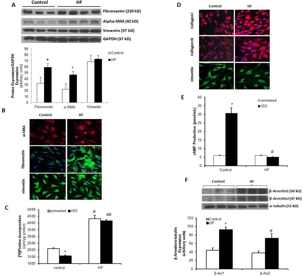
Cardiac fibroblasts (CFs) produce and degrade the myocardial extracellular matrix and are critical in maladaptive ventricular remodeling that can result in heart failure (HF). β-Arrestins are important signaling molecules involved in β-adrenergic receptor (β-AR) desensitization and can also mediate signaling in a G protein-independent fashion. We hypothesize that β-arrestins play an important role in the regulation of adult human CF biology with regard to myofibroblast transformation, increased collagen synthesis, and myocardial fibrosis which are important in the development of HF. β-Arrestin1 & 2 expression is significantly upregulated in adult human CF isolated from failing left ventricles and β-AR signaling is uncoupled with loss of β-agonist-mediated inhibition of collagen synthesis versus normal control CF. Knockdown of either β-arrestin1 or 2 restored β-AR signaling and β-agonist mediated inhibition of collagen synthesis. Overexpression of β-arrestins in normal CF led to a failing phenotype with increased baseline collagen synthesis, impaired β-AR signaling, and loss of β-agonist-mediated inhibition of collagen synthesis. β-Arrestin knockdown in failing CF diminished TGF-β stimulated collagen synthesis and also inhibited ERK phosphorylation. Overexpression of β-arrestins in normal CF increased basal ERK1/2 and Smad2/3 phosphorylation and enhanced TGF-β-stimulated collagen synthesis. This was prevented by pre-treatment with a MEK1/2 inhibitor. Enhanced β-arrestin signaling appears to be deleterious in CF by promoting a pro-fibrotic phenotype via uncoupling of β-AR signaling as well as potentiating ERK and Smad signaling. Targeted inhibition of β-arrestins in CF may represent a therapeutic strategy to prevent maladaptive myocardial fibrosis.

Keywords: Heart failure; Receptors; Signal transduction.


Figures

Figure 1
Figure 1. Heart failure leads to uncoupling of β-adrenergic receptor signaling and up-regulation of β-arrestins
(A) Representative immunoblot (upper panel) showing significant increase in α-SMA and fibronectin in failing (HF) human CF compared with non-failing (control) CF. Vimentin and GAPDH were used as loading controls. Densitometric analysis of α-SMA and fibronectin expression normalized to vimentin and GAPDH is shown below. n=3–4 in each group; *p<0.05 vs. control. (B) Confocal images (40x) of α-SMA and fibronectin immunostaining with red AlexaFluor 594 dye, vimentin is stained with green AlexaFluor 488 dye shown in control and HF cells. Nuclei are stained blue with DAPI. Scale bar=10 µm. (C) Collagen synthesis in control CF and heart failure CF under basal conditions and following ISO (10 µM) treatment. n=3–5 in each group; *p<0.03 vs. control (untreated), #p<0.001 vs. control (untreated), ##p<0.001 vs. control (Iso) and p>0.05 vs. HF (untreated). (D) Confocal images (40x) of collagens types I and III in CF. Nuclei are stained blue with DAPI. Vimentin was used as a fibroblast marker protein visualized with green AlexaFluor 488. Scale bar=10 µm. (E) Basal and ISO-stimulated intracellular cAMP levels in control and HF cardiac fibroblasts. n=4 in each group; *p<0.002 vs. control (untreated) and #p<0.002 vs. control (ISO). (F) Representative immunoblot (upper panel) and densitometric analysis (lower panel) demonstrate a 2.2-fold increase in β-arrestin 1 and a 2-fold increase in β-arrestin 2 in HF compared to control CF. Values are normalized to α-tubulin. n=3–5 in each group; *p<0.002 vs. control, #p<0.05 vs. control.
Figure 2
Figure 2. Inhibition of β-arrestins in failing CF inhibits transformation to myofibroblasts
(A) Failing CF were transfected with siRNA of β-arrestins for 48 hours. Representative immunoblot showing knockdown effects of β-arrestin 1 in HF. The lower panel shows densitometric analysis of β-arrestin 1 expression normalized to GAPDH. n=4 in each group; *p=0.03 vs. scr-siRNA control. (B) Failing CF were transfected with siRNA of β-arrestin 2 for 48 hours. Representative immunoblot showing knockdown effects of β-arrestin 2 in HF. The lower panel shows densitometric analysis of β-arrestin 2 expression normalized to α-tubulin. n=2 in each group; *p<0.05 vs. scr-siRNA control. (C) Representative immunoblot (upper panel) showing significant decrease in α-SMA after β-arrestin knockdown in failing CF when compared with scr-siRNA. GAPDH was used as a loading control. Densitometric analysis of α-SMA and vimentin expression normalized to GAPDH is shown below. n=3 in each group; *p<0.006 vs. scr-siRNA, #p<0.001 vs. scr-siRNA. (D) Confocal images (40x) of scr-siRNA vs. β-Arr1/2 siRNA in failing CF displaying inhibition of α-SMA and fibronectin expression after β-arrestin knockdown. Purity of the cultures was determined by staining for the fibroblast marker vimentin (red). Nuclei were stained blue with DAPI. Scale bar=10 µm.
Figure 3
Figure 3. Knockdown of β-arrestins in HF CF restores isoproterenol (ISO)-stimulated cAMP production and inhibition of collagen synthesis
(A) ISO (10 µM)-stimulated collagen synthesis in HF CF transfected with either β-arrestin 1 siRNA or β-arrestin 2 siRNA. n=4 in each group; *P<0.03 vs. scr-siRNA (ISO) & vs. β-arr1 siRNA (Untreated), **P<0.006 vs. scr-siRNA (ISO) & vs. β-arr2 siRNA (Untreated). (B) Confocal images (40x) of β-Arr1 or 2 siRNA vs. scr-siRNA in HF fibroblasts demonstrating inhibition of collagen I expression with β-Arr1 or 2 knockdown. Nuclei were stained blue with DAPI. Scale bar=10 µm. (C) ISO (10 µM)-stimulated cAMP production in HF CF following β-arrestin 1 or 2 knockdown. n=3 in each group; *p<0.003 vs. scr-siRNA (ISO) & vs. β-arrestin 1 siRNA (Untreated), #p<0.005 vs. scr-siRNA (ISO) & vs. β-arrestin 2 siRNA (Untreated).
Figure 4
Figure 4. Overexpression of β-arrestins in normal cardiac fibroblasts uncouples β-AR signaling and prevents β-agonist-mediated inhibition of collagen synthesis
(A) Normal CF were infected with adenoviruses overexpressing β-arrestin 1 (Ad-β-arr1), β-arrestin 2 (Ad-β-arr2), or control (Ad-GFP) for 48 hours. Representative immunoblot (upper panel) and densitometric analysis (lower panel) shows overexpression of β-arrestin 1 in normal CF. α-tubulin was used as a loading control. n=3 in each group; *P<0.005 vs. Ad-GFP & vs. Ad-β-arr2. (B) Normal CF were infected with adenoviruses overexpressing β-arrestin 1 (Ad-β-arr1) or β-arrestin 2 (Ad-β-arr2) for 48 hours. Representative immunoblot (upper panel) and densitometric analysis (lower panel) shows overexpression of β-arrestin 2 in normal CF. α-tubulin was used as a loading control. n=2–4 in each group; *P<0.02 vs. Ad-GFP. (C) Collagen synthesis assessed by [3H]proline incorporation under basal conditions and in response to ISO (10µM) in the presence of 2.5% FBS. n=3–4 in each group; *p<0.01 vs. Ad-GFP (ISO), #p<0.05 vs. Ad-GFP (ISO). (D) Representative immunoblot (upper panel) and densitometric analysis (lower panel) of Collagen I expression in normal CF with β-arrestin overexpression or Ad-GFP control. α-tubulin was used as a loading control. n=3 in each group; *p<0.005 vs. Ad-GFP (untreated), #p<0.005 vs. Ad-GFP (ISO). (E) Confocal images (40x) of collagen I in normal CF with either Ad-β-arrestin 1/2 or Ad-GFP with and without ISO (10 µM) treatment. Nuclei were stained blue with DAPI. Scale bar=10 µm. (F) Adenoviral-mediated overexpression of β-arrestins in normal CF leads to impaired basal and ISO (10µM)-stimulated cAMP production n=3–4; *p<0.03 vs. Ad-GFP (untreated), #p<0.01 vs. Ad-GFP (ISO).
Figure 5
Figure 5. Inhibition of β-arrestin expression abolishes TGF-β-induced collagen synthesis and ERK1/2 phosphorylation in failing CF
(A) HF CF were transfected with β-arrestin siRNA and treated with 0.5, 1 and 10 ng/mL of TGF-β for 24 hours. Dose-response curves of TGF-β-stimulated collagen synthesis in HF CF transfected with β-arrestin siRNA. n=4 in each group; *p<0.001 (scr siRNA) vs. β-Arr1 siRNA & vs. β-Arr2 siRNA. (B) HF CF transfected with β-arrestin 1 or 2 siRNA were treated with 10ng/mL of TGF-β for 0, 5, 10, 20, 30, or 60 minutes. Representative immunoblot showing time course of TGF-β-stimulated p-ERK1/2 expression. (C) Densitometric analysis showing decreased p-ERK1/2 expression in β-arrestin knockdown HF CF vs. scr-siRNA control. p-ERK1/2 was normalized to t-ERK1/2. n=4 in each group; *p<0.0001 vs. scr-siRNA + Untreated (0min). (D) Confocal images (40x) of p-ERK1/2 stained green with FITC in HF CF transfected with either β-arrestin siRNA or scr-siRNA with TGF-β (10 ng/mL) treatment at different time points. Nuclei were stained blue with DAPI. Scale bar=10 µm.
Figure 6
Figure 6. TGF-β stimulated collagen synthesis is enhanced by overexpression of β-arrestins in normal CF and is ERK-dependent
(A) Normal CF infected with adenoviruses overexpressing β-arrestin 1 (Ad-β-arr1), β-arrestin 2 (Ad-β-arr2), or control (Ad-GFP) for 48 hours. Dose-response curves of TGF-β-stimulated (0.5, 1 and 10 ng/mL) collagen synthesis in normal CF. n=3–8 in each group; *p<0.03 (Ad-β-Arr1) vs. Ad-GFP, #p<0.05 (Ad-β-Arr2) vs. Ad-GFP. (B) Representative immunoblot shows p-ERK1/2 expression is upregulated by overexpression of β-arrestins in normal CF. p-ERK1/2 was normalized to t-ERK1/2. n=3 in each group; *p<0.003 vs. Ad-GFP, #p<0.01 vs. Ad-GFP. (C) Collagen synthesis in response to TGF-β stimulation (10ng/mL) under 2.5% serum conditions, or pre-treated with PD98059 (10 µM) or UO126 (10 µM) for 20 minutes prior to 48 hours of stimulation with TGF-β (10ng/mL). n=4 in each group; *p<0.001 vs. Ctrl, **p<0.001 vs. TGF-β, #p<0.001 vs. Ad-GFP (Ctrl).
Figure 7
Figure 7. β-arrestin-induced Smad2/3 phosphorylation is ERK-dependent and β-arrestin-mediated collagen synthesis is Smad-dependent
(A) Representative immunoblot (upper panel) and densitometric analysis (lower panel) show TGF-β-stimulated Smad2/3 phosphorylation is regulated by the ERK signaling pathway. p-Smad2/3 was normalized to t-Smad2/3. n=3 in each group; *P<0.01 vs. Ad-GFP. (B) Confocal images (40x) of p-Smad2/3 in normal control CF infected with either Ad-β-arr1 or 2 or Ad-GFP with or without TGF-β (10 ng/mL) treatment. MEK inhibitor U0126 pre-treatment inhibits TGF-β-stimulated p-Smad2/3 translocation into nuclei. Scale bar=10 µm. (C) Normal CF were transfected with siRNA for Smad2/3 for 48 hours. Representative immunoblot showing effective knockdown of Smad2/3 in Normal CF. Smad2/3 was normalized to GAPDH. n=3 in each group. (D) Collagen synthesis in response to TGF-β stimulation (10ng/mL) under 2.5% serum conditions. Normal CF were infected with Ad-β-arrestin 1 or 2, then transfected with Smad2/3 siRNA. n=3–6 in each group; *p<0.02 vs. scr-siRNA (untreated), #p<0.01 vs. scr-siRNA (TGF-β).

References

    1. Swynghedauw B. Molecular mechanisms of myocardial remodeling. Physiol Rev. 1999;79:215–262. - PubMed
    1. Chatterjee K, Massie B. Systolic and diastolic heart failure: Differences and similarties. J Card Fail. 2007;13:569–576. - PubMed
    1. Konstam MA, Udelson JE, Anand IS, Cohn JN. Ventricular remodeling in heart failure: A credible surrogate endpoint. J Card Fail. 2003;9:350–353. - PubMed
    1. Camelliti P, Borg TK, Kohl P. Structural and functional characterization of cardiac fibroblasts. Cardiovasc Res. 2005;65:40–51. - PubMed
    1. van den Borne SWM, Diez J, Blankesteijn WM, Verjans J, Hofstra L, Narula J. Myocardial remodeling after infarction: the role of myofibroblasts. Nat Rev Cardiol. 2010;7:30–37. - PubMed

Publication types

MeSH terms