. 2015 Nov;220(6):3369-84.
doi: 10.1007/s00429-014-0861-2. Epub 2014 Aug 19.

Contralateral cerebello-thalamo-cortical pathways with prominent involvement of associative areas in humans in vivo

Affiliations

Contralateral cerebello-thalamo-cortical pathways with prominent involvement of associative areas in humans in vivo

Fulvia Palesi et al. Brain Struct Funct. 2015 Nov.

Abstract

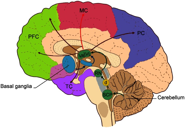
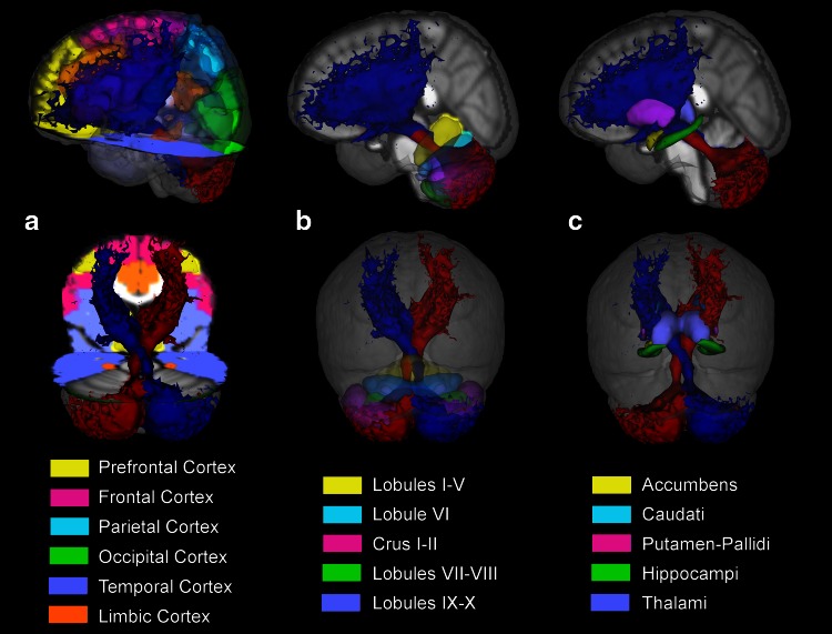
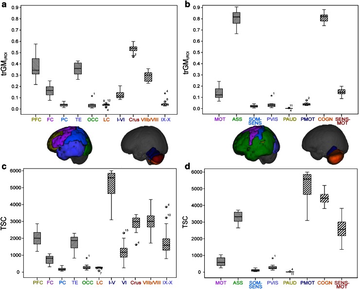
In addition to motor functions, it has become clear that in humans the cerebellum plays a significant role in cognition too, through connections with associative areas in the cerebral cortex. Classical anatomy indicates that neo-cerebellar regions are connected with the contralateral cerebral cortex through the dentate nucleus, superior cerebellar peduncle, red nucleus and ventrolateral anterior nucleus of the thalamus. The anatomical existence of these connections has been demonstrated using virus retrograde transport techniques in monkeys and rats ex vivo. In this study, using advanced diffusion MRI tractography we show that it is possible to calculate streamlines to reconstruct the pathway connecting the cerebellar cortex with contralateral cerebral cortex in humans in vivo. Corresponding areas of the cerebellar and cerebral cortex encompassed similar proportion (about 80%) of the tract, suggesting that the majority of streamlines passing through the superior cerebellar peduncle connect the cerebellar hemispheres through the ventrolateral thalamus with contralateral associative areas. This result demonstrates that this kind of tractography is a useful tool to map connections between the cerebellum and the cerebral cortex and moreover could be used to support specific theories about the abnormal communication along these pathways in cognitive dysfunctions in pathologies ranging from dyslexia to autism.

Keywords: Cerebellum; Cerebral cortex; Diffusion MRI; MRI tractography.


Figures

Fig. 1
Fig. 1
The most important connections in the cerebello-cortical circuit. Projections from the basal ganglia (through the subthalamic nucleus, STN) go mainly to the thalamic nuclei (VA/VL). The cerebellum sends its output through the superior cerebellar peduncle (SCP), the contralateral red nucleus (RN), and VA/VL of the thalamus to various cerebral areas including the motor cortex (MC), the prefrontal cortex (PFC), the parietal cortex (PC), and the temporal cortex (TC). The decussation (d) of the cerebello-thalamo-cortical pathway is indicated by the yellow circle. Modified from D’Angelo and Casali (2013)
Fig. 2
Fig. 2
The two tractography metrics: cROItr and trGMcROI. Each different colour (green, red, yellow) and capital letter represents a different cortical parcellation (cROI) while each lowercase letter represents the cortical region reached by the tract for each cROI
Fig. 3
Fig. 3
Example of cerebello-thalamo-cortical pathway from a representative subject. This is a 2D rendering of streamlines extending over a volume of 5 mm, but mapped to a section of 1 mm thick. The same seed ROI (ac) was placed on left superior cerebellar peduncle. a The tract was reconstructed using DTI and streamline tractography. No target ROI was drawn. b The tract was reconstructed using a combination of the CSD algorithm and probabilistic tractography. No target ROI was drawn. c The tract was reconstructed as in b with a target ROI drawn on the whole contralateral red nucleus. d Details of the fibre-orientation distribution (FOD) within the decussation region. e Details of the FOD and tract within the decussation region
Fig. 4
Fig. 4
2D rendering of both left and right cerebello-thalamo-cortical pathways from a representative subject. a The tracts are colour-coded by direction to follow the anatomy and the directionality of both left and right tracts. b The tracts are reported using a single solid colour for each tract to distinguish the streamlines from the left (red) and right (blue) pathways
Fig. 5
Fig. 5
Tridimensional view of the average cerebello-thalamo-cortical pathway across all subjects in MNI space. Cerebral (a), cerebellar (b) and deep grey matter (c) atlases are overlaid to assist visualization of cerebellar connections. a Distribution of left (red) and right (blue) tracts in the cerebral cortex: the reconstructed tracts reach the prefrontal (yellow), frontal (fuchsia) and temporal (violet) cortices with greater density of streamlines. b Streamlines distribution in the cerebellar cortex: the lateral Crus I–II (fuchsia) and the lateral lobules VIIb/VIII (green) are showing the greatest density of tracts. c Streamlines distribution of deep grey matter nuclei: the thalami (violet), the caudate (light blue) and the putamen (fuchsia) show the greatest trGMcROI
Fig. 6
Fig. 6
Extension of the left cerebello-thalamo-cortical pathway overlapped to the parcellated thalami in a representative subject. L indicates the left side of the brain. a 2D rendering: the highest density of streamlines is seen in the VA and VL nuclei of the thalamus, which correspond to areas principally connected with prefrontal (yellow) and frontal (orange and blue) cortices. b Tridimensional representation of the tract: the VA and VL nuclei of the right thalamus (yellow, orange and blue) are hidden from the tract
Fig. 7
Fig. 7
Histograms of mean values of trGMcROI and TSC for each region of the cerebral cortex and of the cerebellar cortex. PFC prefrontal cortex, FC frontal cortex, PC parietal cortex, TE temporal cortex, OCC occipital cortex, LC limbic cortex, MOT motor, ASS associative, SOM-SEN somatosensory, PVIS primary visual, PAUD primary auditory, PMOT primary motor, COGN cognitive/sensory, SEN-MOT sensory motor. a trGMcROI values of all cerebral and cerebellar regions created on anatomical bases. PFC and Lateral Crus I–II have the highest values in cerebrum and cerebellum, respectively. b TSC values of all regions created on anatomical bases. PFC and lobules I–V have the highest values in cerebrum and cerebellum, respectively. c trGMcROI values of all regions created on functional bases. ASS and COGN areas have the highest values in cerebrum and cerebellum, respectively. d TSC values of all regions created on functional bases. ASS and PMOT areas have the highest values in cerebrum and cerebellum, respectively

References

    1. Akhlaghi H, Yu J, Corben L et al (2013) Cognitive deficits in friedreich ataxia correlate with micro-structural changes in dentatorubral tract. Cerebellum. doi:10.1007/s12311-013-0525-4 - PubMed
    1. Alexander AL, Hasan KM, Lazar M, et al. Analysis of partial volume effects in diffusion-tensor MRI. Magn Reson Med. 2001;45:770–780. doi: 10.1002/mrm.1105. - DOI - PubMed
    1. Alexander DC, Barker GJ, Arridge SR. Detection and modeling of non-Gaussian apparent diffusion coefficient profiles in human brain data. Magn Reson Med. 2002;48:331–340. doi: 10.1002/mrm.10209. - DOI - PubMed
    1. Amaral DG. The functional organization of perception and movement. In: Kandel ER, Schwartz JH, Jessell TM, editors. Princ. Neural Sci. 4. New York: McGraw-Hill; 2000. pp. 337–348.
    1. Amodio DM, Frith CD. Meeting of minds: the medial frontal cortex and social cognition. Nat Rev Neurosci. 2006;7:268–277. doi: 10.1038/nrn1884. - DOI - PubMed

Publication types

LinkOut - more resources