. 2014 Nov;196(21):3746-55.
doi: 10.1128/JB.01799-14. Epub 2014 Aug 18.

HilD induces expression of Salmonella pathogenicity island 2 genes by displacing the global negative regulator H-NS from ssrAB

Affiliations

HilD induces expression of Salmonella pathogenicity island 2 genes by displacing the global negative regulator H-NS from ssrAB

Luary C Martínez et al. J Bacteriol. 2014 Nov.

Abstract

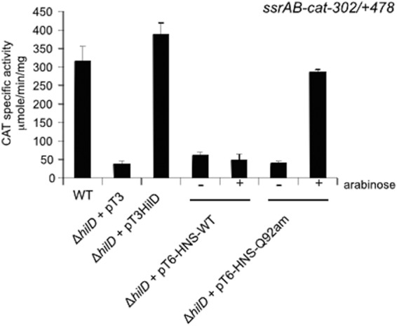
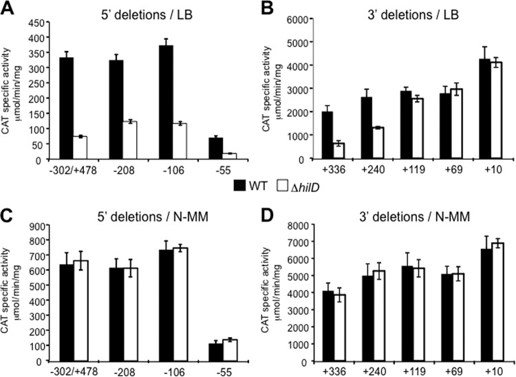
Salmonella pathogenicity islands 1 and 2 (SPI-1 and SPI-2) have essential roles in the pathogenesis of Salmonella enterica. Previously, we reported transcriptional cross talk between SPI-1 and SPI-2 when the SPI-1 regulator HilD induces expression of the SsrA/B two-component system, the central positive regulator of SPI-2, during the growth of Salmonella to late stationary phase in LB rich medium. Here, we further define the mechanism of the HilD-mediated expression of ssrAB. Expression analysis of cat transcriptional fusions containing different regions of ssrAB revealed the presence of negative regulatory sequences located downstream of the ssrAB promoter. In the absence of these negative cis elements, ssrAB was expressed in a HilD-independent manner and was no longer repressed by the global regulator H-NS. Consistently, when the activity of H-NS was inactivated, the expression of ssrAB also became independent of HilD. Furthermore, electrophoretic mobility shift assays showed that both HilD and H-NS bind to the ssrAB region containing the repressing sequences. Moreover, HilD was able to displace H-NS bound to this region, whereas H-NS did not displace HilD. Our results support a model indicating that HilD displaces H-NS from a region downstream of the promoter of ssrAB by binding to sites overlapping or close to those sites bound by H-NS, which leads to the expression of ssrAB. Although the role of HilD as an antagonist of H-NS has been reported before for other genes, this is the first study showing that HilD is able to effectively displace H-NS from the promoter of one of its target genes.


Figures

FIG 1
FIG 1
Schematic representation of the ssrAB loci and the ssrAB-cat transcriptional fusions used in this work. The transcriptional start site (+1) of ssrAB is indicated by a bent arrow. The positions spanning the region between ssaB and ssrAB are shown. The transcriptional fusions are represented as lines with a short black arrow at their 3′ ends, indicating the respective ssrAB region (lines) and the cat reporter gene (arrows). The positions spanning the ssrAB fragment carried by each fusion, as well as the name of each fusion, are shown. Fusions are grouped with regard to 5′ or 3′ deletions with respect to the ssrAB-cat−302/+478 fusion. All positions are indicated with respect to the transcriptional start site of ssrAB.
FIG 2
FIG 2
HilD antagonizes the H-NS-mediated repression exerted on ssrAB. Expression of the ssrAB-cat−302/+478 transcriptional fusion, carried by plasmid pssrAB-cat−302/+478, was tested in the WT S. Typhimurium strain and its isogenic ΔhilD mutant containing the vector pMPM-T3 or the plasmid pT3-HilD1, which expresses HilD, as well as in the ΔhilD mutant containing the plasmid pT6-HNS-WT or pT6-HNS-Q92am, which express WT H-NS or a dominant negative C-terminally truncated form of H-NS (H-NSQ92am), respectively, from an arabinose-inducible promoter. CAT-specific activity was determined from samples collected from bacterial cultures grown for 10 h in LB medium at 37°C. Expression of WT H-NS or H-NSQ92am from plasmids pT6-HNS-WT and pT6-HNS-Q92am, respectively, was induced by adding 0.1% l-arabinose to the medium (+arabinose). The data are the averages of results of three independent experiments performed in duplicate. Bars represent the standard deviations.
FIG 3
FIG 3
Analysis of the cis-acting sequences required for the HilD-mediated regulation of ssrAB. Expression of the ssrAB-cat−302/+478, ssrAB-cat−208, ssrAB-cat−106, ssrAB-cat−55, ssrAB-cat+336, ssrAB-cat+240, ssrAB-cat+119, ssrAB-cat+69, and ssrAB-cat+10 transcriptional fusions contained in plasmids pssrAB-cat−302/+478, pssrAB-cat−208, pssrAB-cat−106, pssrAB-cat−55, pssrAB-cat+336, pssrAB-cat+240, pssrAB-cat+119, pssrAB-cat+69, and pssrAB-cat+10, respectively, was tested in the WT S. Typhimurium strain and its isogenic ΔhilD mutant. CAT-specific activity was determined from samples collected from bacterial cultures grown in LB medium (A and B) or N-MM (C and D) at 37°C for 10 h or 16 h, respectively. The data are the averages of results from three independent experiments performed in duplicate. Bars represent the standard deviations.
FIG 4
FIG 4
Analysis of the cis-acting sequences required for the H-NS-mediated repression of ssrAB. Expression of the ssrAB-cat−302/+478, ssrAB-cat−208, ssrAB-cat−106, ssrAB-cat−55, ssrAB-cat+336, ssrAB-cat+240, ssrAB-cat+119, ssrAB-cat+69, and ssrAB-cat+10 transcriptional fusions contained in plasmids pssrAB-cat−302/+478, pssrAB-cat−208, pssrAB-cat−106, pssrAB-cat−55, pssrAB-cat+336, pssrAB-cat+240, pssrAB-cat+119, pssrAB-cat+69, and pssrAB-cat+10, respectively, was tested in the S. Typhimurium ΔhilD mutant containing the plasmid pT6-HNS-Q92am, which expresses a dominant negative C-terminally truncated form of H-NS (H-NSQ92am) from an arabinose-inducible promoter. (A and B) Results for the 5′- and 3′-deletion fusions, respectively. CAT-specific activity was determined from samples collected from bacterial cultures grown for 10 h in LB medium at 37°C. Expression of H-NSQ92am from pT6-HNS-Q92am was induced by adding 0.1% l-arabinose to the medium (+ arabinose). The data are the averages of results from three independent experiments performed in duplicate. Bars represent the standard deviations.
FIG 5
FIG 5
HilD and H-NS bind to the same regions of ssrAB. (A) Schematic representation of the ssrAB loci and the five ∼200-bp overlapping DNA fragments (1A to 1E) spanning the −302/+478 region that were used in the EMSAs. Important positions, with respect to the transcriptional start site of ssrAB, are indicated. The two different regions (−111/+37 and +136/+287) bound by HilD and H-NS, deduced from the EMSAs with the five overlapping DNA fragments, are indicated by two gray-filled boxes inside the dashed-line box representing the entire region (−111/+287) bound by these proteins. (B) EMSAs. The ssrAB DNA fragments 1A to 1E were incubated with increasing concentrations of purified MBP-HilD (0, 0.2, 0.3, 0.4, and 0.5 μM) or H-NS–FH (0, 0.3, 0.4, 0.5, and 0.6 μM). A fragment containing the regulatory region of sigD, a gene that is not regulated directly by HilD or H-NS, was used as a negative control. The DNA-protein complexes, which are indicated by an asterisk, were resolved in a nondenaturing 6% polyacrylamide gel and stained with ethidium bromide.
FIG 6
FIG 6
HilD displaces H-NS from ssrAB. Shown are the results of competitive EMSAs between H-NS, which was added first, and HilD (A) or between HilD, which was added first, and H-NS (B) in the −302/+478 region of ssrAB. The upper panels show the ethidium bromide-stained gels, and the middle and lower panels show the immunoblot detection of H-NS–FH and MBP-HilD from the DNA-protein complexes, respectively, obtained from different membranes. (A) H-NS–FH was added at 0.6 μM (lanes 3 to 7), and MBP-HilD was added at 0.2, 0.3, 0.4, and 0.5 μM (lanes 4 to 7, respectively). No protein was added in lane 1, and MBP-HilD was added at 0.5 μM in lane 2. (B) MBP-HilD was added at 0.5 μM (lanes 3 to 7), and H-NS–FH was added at 0.3, 0.4, 0.5, and 0.6 μM (lanes 4 to 7). No protein was added in lane 1, and H-NS–FH was added at 0.6 μM in lane 2. Similar results were obtained from three different experiments.
FIG 7
FIG 7
Model for the HilD- and H-NS-mediated regulation of ssrAB. H-NS represses ssrAB by binding to multiple sites located in a region spanning the promoter and downstream sequence, which may form an H-NS filament complex that blocks the access of the RNA polymerase to the ssrAB promoter. HilD binds to sites overlapping or close to most of the sites bound by H-NS and thus displaces H-NS from most of its binding sites, which disrupts the H-NS nucleoprotein complex, thus leading to the expression of ssrAB. The regions required for the regulation of ssrAB by HilD and H-NS, defined in this study by expression and binding assays, are shown.

References

    1. Schmidt H, Hensel M. 2004. Pathogenicity islands in bacterial pathogenesis. Clin. Microbiol. Rev. 17:14–56. 10.1128/CMR.17.1.14-56.2004. - DOI - PMC - PubMed
    1. Fàbrega A, Vila J. 2013. Salmonella enterica serovar Typhimurium skills to succeed in the host: virulence and regulation. Clin. Microbiol. Rev. 26:308–341. 10.1128/CMR.00066-12. - DOI - PMC - PubMed
    1. Hansen-Wester I, Hensel M. 2001. Salmonella pathogenicity islands encoding type III secretion systems. Microbes Infect. 3:549–559. 10.1016/S1286-4579(01)01411-3. - DOI - PubMed
    1. Haraga A, Ohlson MB, Miller SI. 2008. Salmonellae interplay with host cells. Nat. Rev. Microbiol. 6:53–66. 10.1038/nrmicro1788. - DOI - PubMed
    1. Moest TP, Meresse S. 2013. Salmonella T3SSs: successful mission of the secret(ion) agents. Curr. Opin. Microbiol. 16:38–44. 10.1016/j.mib.2012.11.006. - DOI - PubMed

Publication types

MeSH terms

LinkOut - more resources