. 2015 Jan;9(1):44-57.
doi: 10.1016/j.molonc.2014.07.013. Epub 2014 Jul 29.

Infiltrating T cells promote prostate cancer metastasis via modulation of FGF11→miRNA-541→androgen receptor (AR)→MMP9 signaling

Affiliations

Infiltrating T cells promote prostate cancer metastasis via modulation of FGF11→miRNA-541→androgen receptor (AR)→MMP9 signaling

Shuai Hu et al. Mol Oncol. 2015 Jan.

Erratum in

Abstract

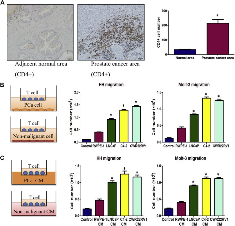
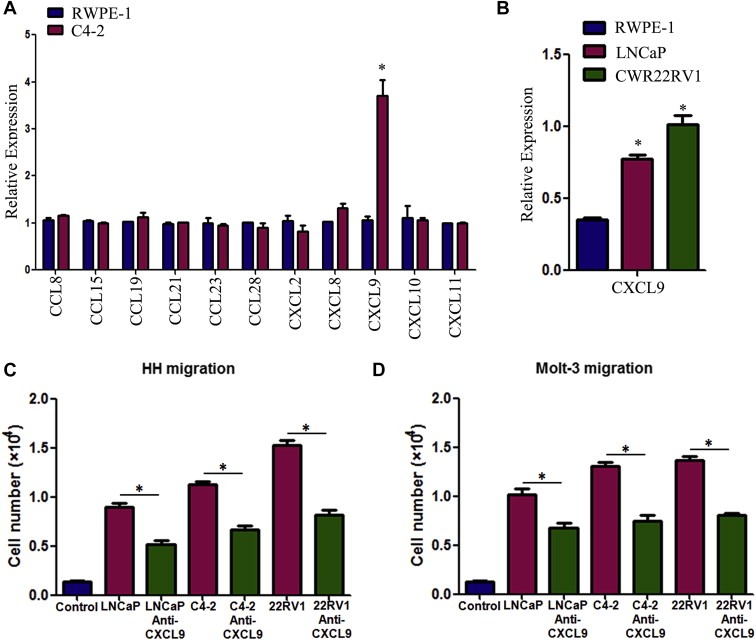
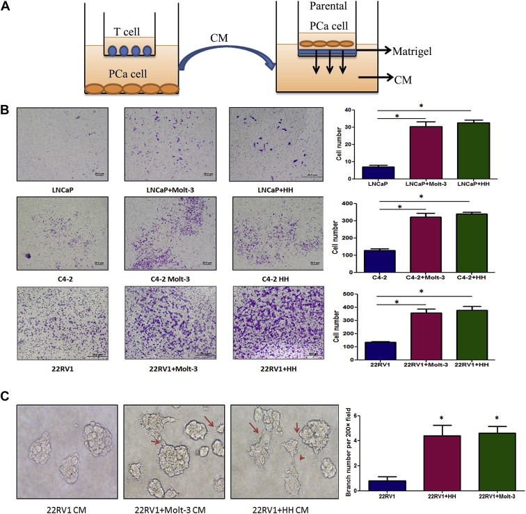
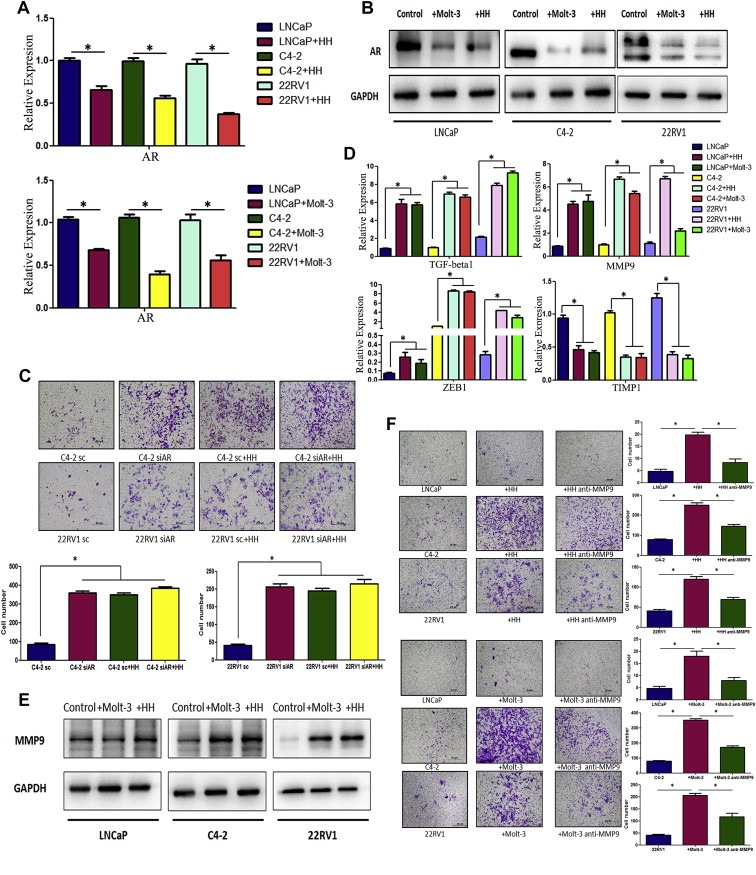
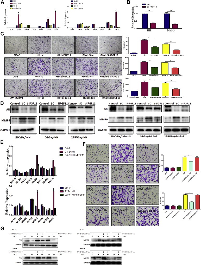
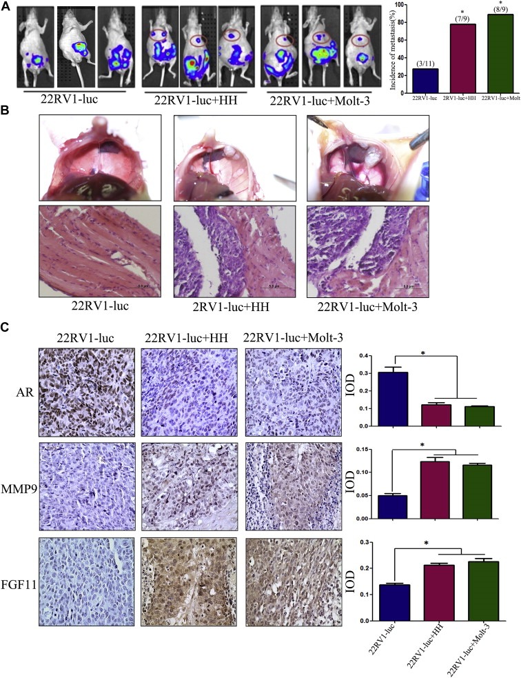
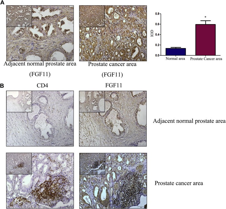
Early clinical studies suggested infiltrating T cells might be associated with poor outcomes in prostate cancer (PCa) patients. The detailed mechanisms how T cells contribute to PCa progression, however, remained unclear. Here, we found PCa cells have a better capacity to recruit more CD4(+) T cells than the surrounding normal prostate cells via secreting more chemokines-CXCL9. The consequences of more recruited CD4(+) T cells to PCa might then lead to enhance PCa cell invasion. Mechanism dissection revealed that infiltrating CD4(+) T cells might function through the modulation of FGF11→miRNA-541 signals to suppress PCa androgen receptor (AR) signals. The suppressed AR signals might then alter the MMP9 signals to promote the PCa cell invasion. Importantly, suppressed AR signals via AR-siRNA or anti-androgen Enzalutamide in PCa cells also enhanced the recruitment of T cells and the consequences of this positive feed back regulation could then enhance the PCa cell invasion. Targeting these newly identified signals via FGF11-siRNA, miRNA-541 inhibitor or MMP9 inhibitor all led to partially reverse the enhanced PCa cell invasion. Results from in vivo mouse models also confirmed the in vitro cell lines in co-culture studies. Together, these results concluded that infiltrating CD4(+) T cells could promote PCa metastasis via modulation of FGF11→miRNA-541→AR→MMP9 signaling. Targeting these newly identified signals may provide us a new potential therapeutic approach to better battle PCa metastasis.

Keywords: Androgen receptor; Prostate cancer; T cells; Tumor metastasis; Tumor microenvironment.


Figures

Figure 1
Figure 1
Prostate cancer tissues recruit more T cells. A. Clinical specimen IHC staining from 20 prostate cancer patients showed that more CD4(+) T cells infiltrate surrounding the prostate cancer area than in the adjacent normal prostate area, scale bar: 20 μm, *P < 0.05. B. Transwell T cell migration assay. 1 × 105 of PCa cells or non‐malignant prostate epithelial cells were plated into the lower chambers of the transwells. 1 × 105 of CD4(+) T cells, HH cells or Molt‐3 cells, were plated onto the upper chamber with 5 μm pore polycarbonate membrane for T cell migration assay. The cells migrated into the lower chamber media were collected after 6 h and counted by the Bio‐Rad TC10 automatic cell counter. Compared to the prostate normal epithelial cell line RWPE‐1, LNCaP, C4‐2 and CWR22RV1 recruited more HH and Molt‐3 (*P < 0.05). C. When PCa or RWPE‐1 conditioned media (CM) was added into the lower chamber, PCa cell CM could attract more HH or Molt‐3 cells compared with RWPE‐1 cells CM (*P < 0.05).
Figure 2
Figure 2
Reverse of T cell recruitment by blocking the CXCL9 with neutralizing antibody. A. Q‐PCR screening of a panel of cytokine factors that could be responsible for PCa cell promoted T cell migration. Compared to the RWPE‐1 cells, C4‐2 cells can express more CXCL9 (*P < 0.05). B. Higher expression of CXCL9 mRNA was confirmed in LNCaP and CWR22RV1 (22RV1) cell lines using q‐PCR (*P < 0.05). C&D. The PCa cell‐promoted HH and Molt‐3 migrations were partly blocked after adding CXCL9 neutralizing antibody (4 μg/ml) into PCa CM in bottom chambers (*P < 0.05).
Figure 3
Figure 3
Co‐culture of T cells promoted PCa invasion. A. CMs collected from PCa/T cells co‐culture could promote PCa invasiveness using in vitro Matrigel invasion assay. The PCa/T cells were co‐cultured in 0.4 μM transwell plate for 2 days. The CM or control media were collected, diluted 1:1 with 10% FBS RPMI media, and placed into the lower chamber of transwells. The 1 × 105 PCa cells (without any pre‐treatment) were plated onto upper insert chamber pre‐coated with Matrigel for 36 h invasion assay. B. Toluidine blue staining results showed the PCa/T cells co‐cultured CM can promote PCa cell invasion. Invasion assay has been performed on 3 different PCa cell lines, LNCaP, C4‐2 and CWR22RV1 (22RV1) and 2 different T cells, Molt‐3 and HH (*P < 0.05). C. 3D invasion assay showed that more acini‐like structures formed when treating the parental CWR22RV1 cells with CWR22RV1/T cell co‐culture CM (200×) (*P < 0.05).
Figure 4
Figure 4
T cells can down‐regulate AR expression and up‐regulate MMP9 expression of PCa cells after co‐culture. A. Q‐PCR detection showed AR mRNA was down‐regulated in PCa cells, LNCaP, C4‐2 and CWR22RV1 (22RV1), after co‐culture with HH or Molt‐3 cells for 2 days (*P < 0.05). B. Western‐blot results showed AR expression of LNCaP, C4‐2 and 22RV1 cells decreased after co‐culture with HH or Molt‐3 cells for 2 days. C. T cell co‐culture could not effectively change invasion in PCa cells with AR knockdown by siRNA. We used lentiviral AR‐siRNA to knock‐down AR expression in PCa. T cells co‐culture failed to promote invasion of PCa cell with AR‐siRNA compared with PCa cell with control si‐Luc (*P < 0.05). D. Four metastasis–related genes were detected by q‐PCR. TGF‐beta1, MMP9 and ZEB1 mRNA level were up‐regulated in PCa cells after co‐culture with T cells, however the MMP9‐inhibitor gene, TIMP1, decreased in PCa cells (*P < 0.05). E. MMP9 expressions of LNCaP, C4‐2 and 22RV1 cells are up‐regulated after co‐culture with HH or Molt‐3 cells using Western‐blot assay (*P < 0.05). F. Blocking MMP9 by MMP9 inhibitor can partly reverse T cell‐promoted PCa invasion (*P < 0.05).
Figure 5
Figure 5
Down‐regulation of AR in PCa cells by T cells secreted FGF11. A. Results showed FGF11 expression is selectively and significantly increased in T cells after co‐culture with PCa cells for 2 days. The data have been validated in 2 different T cell lines co‐cultured with C4‐2 cells (*P < 0.05). B. Validation of FGF11‐siRNA knockdown efficiency in 2 different T cell lines (*P < 0.05). C. T cell promotion of PCa cell invasion can be partly reversed by FGF11 knock‐down in 2 T cell lines. The results have been validated in 2 different T cells and 3 PCa cells, LNCaP, C4‐2, and CWR22RV1 (22RV1) cells (*P < 0.05). D. Knockdown of FGF11 in T cells can partially reverse the T cells‐mediated AR down‐regulation and MMP9 up‐regulation. Results have been validated in the 2 different T cells and 3 PCa cells. E. Q‐PCR results showed that miRNA‐541 (Mir541) and miRNA‐876 (Mir876) stably reduced in PCa cells after knockdown of FGF11 in T cells co‐culture with PCa cells (*P < 0.05). F. MiRNA‐541 inhibitor can partly reverse the HH cell‐enhanced PCa cell invasion (*P < 0.05). G. Inhibition of miRNA‐541 (but not miRNA‐876) can reverse the HH cells‐mediated AR down‐regulation and MMP9 up‐regulation.
Figure 6
Figure 6
Induction of PCa metastasis by T cells in orthotopic PCa model. A. CWR22RV1‐luc (22RV1‐luc) cells (1 × 106) were mixed with HH or Molt‐3 (1 × 105) and orthotopically implanted into the anterior prostates of nude mice. After 6 weeks, the PCa growth and metastases were evaluated by the IVIS system. The 22RV1 cells co‐injected with T cells showed significant increase of the distant metastatic tumors in diaphragm, compared to the 22RV1 single injection mice (*P < 0.05). B. The mice were then sacrificed (6 week after implantation), the metastatic tumors observed on the diaphragm were confirmed by histology H&E staining. C. IHC staining confirmed T cells co‐implanted 22RV1 tumors have a reduced AR, increased MMP9 and FGF11 expression (400×) (*P < 0.05).
Figure 7
Figure 7
Expression of FGF11 in clinical samples. A. IHC staining for FGF11 in tissue specimens from 20 PCa patients. Compared to adjacent normal area (left panel), FGF11 expression was higher in the PCa area (right panel) (scale bar: 50 μm, the scale bar of upper left corners: 100 μm, *P < 0.05). B. Serial paraffin sections were used to detect the distribution of CD4(+) T cells and expression of FGF11 (Upper panels, adjacent normal area; lower panels, PCa area. Left panels CD4(+) and right labels FGF11, scale bar: 50 μm. The scale bar of upper left corners: 100 μm).

References

    1. Bartel, D.P. , 2009. MicroRNAs: target recognition and regulatory functions. Cell. 136, 215–233. - PMC - PubMed
    1. Bromwich, E.J. , McArdle, P.A. , Canna, K. , McMillan, D.C. , McNicol, A.M. , Brown, M. , Aitchison, M. , 2003. The relationship between T-lymphocyte infiltration, stage, tumour grade and survival in patients undergoing curative surgery for renal cell cancer. Br. J. Cancer. 89, 1906–1908. - PMC - PubMed
    1. Chang, C. , Lee, S.O. , Yeh, S. , Chang, T.M. , 2013. Androgen receptor (AR) differential roles in hormone-related tumors including prostate, bladder, kidney, lung, breast and liver. Oncogene. 33, 3225–3234. - PubMed
    1. Chang, C.S. , Kokontis, J. , Liao, S.T. , 1988. Molecular cloning of human and rat complementary DNA encoding androgen receptors. Science. 240, 324–326. - PubMed
    1. Ebelt, K. , Babaryka, G. , Figel, A.M. , Pohla, H. , Buchner, A. , Stief, C.G. , Eisenmenger, W. , Kirchner, T. , Schendel, D.J. , Noessner, E. , 2008. Dominance of CD4+ lymphocytic infiltrates with disturbed effector cell characteristics in the tumor microenvironment of prostate carcinoma. Prostate. 68, 1–10. - PubMed

Publication types

MeSH terms