

Multi-centre analysis of incidental findings on low-resolution CT attenuation correction images
- PMID: 25135310
- PMCID: PMC4170858
- DOI: 10.1259/bjr.20130701
Multi-centre analysis of incidental findings on low-resolution CT attenuation correction images
Abstract
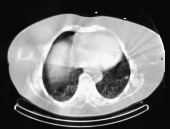
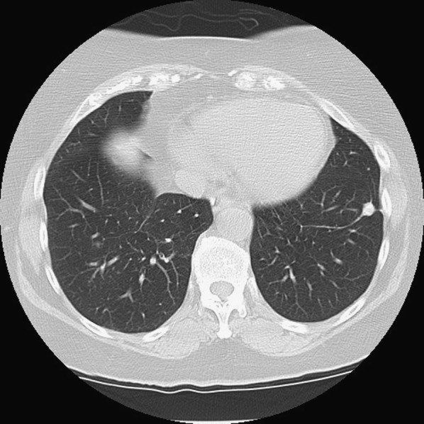
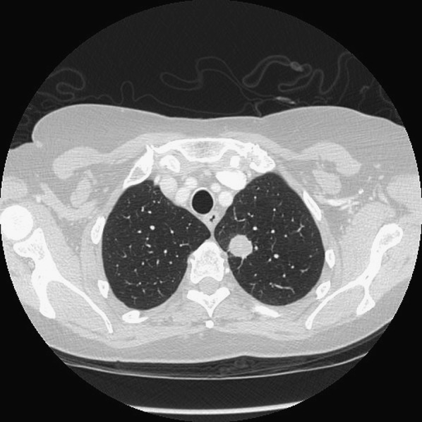
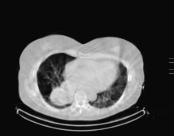
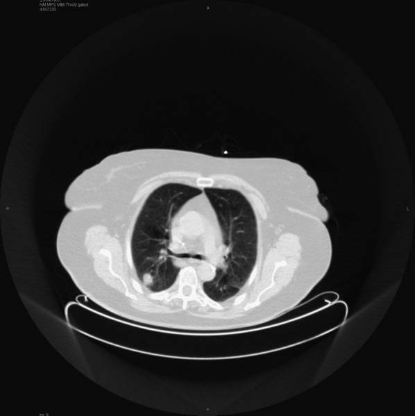
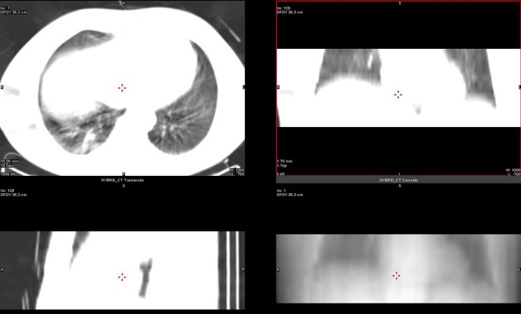
Objective: To review new incidental findings detected on low-resolution CT attenuation correction (CTAC) images acquired during single-photon emission CT (SPECT-CT) myocardial perfusion imaging (MPI) and to determine whether the CTAC images had diagnostic value and warrant reporting.
Methods: A multicentre study was performed in four UK nuclear medicine departments. CTAC images acquired as part of MPI performed using SPECT were evaluated to identify incidental findings. New findings considered to be clinically significant were evaluated further. Positive predictive value (PPV) was determined at the time of definitive diagnosis.
Results: Of 1819 patients studied, 497 (27.3%) had a positive CTAC finding. 51 (2.8%) patients had findings that were clinically significant at the time of the CTAC report and had not been previously diagnosed. Only four (0.2%) of these were potentially detrimental to patient outcome.
Conclusion: One centre had a PPV of 0%, and the study suggests that these CTAC images should not be reported. Two centres with more modern equipment had low PPVs of 0% and 6%, respectively, and further research is suggested prior to drawing a conclusion. The centre with best quality CT had a PPV of 67%, and the study suggests that CTAC images from this equipment should be reported.
Advances in knowledge: This study is unique compared with previous studies that have reported only the potential to identify incidental findings on low-resolution CT images. This study both identifies and evaluates new clinically significant incidental findings, and it demonstrates that the benefit of reporting the CTAC images depends on the type of equipment used.
Figures
Similar articles
-
Multicentre analysis of incidental findings on low-resolution CT attenuation correction images: an extended study.Br J Radiol. 2015;88(1056):20150555. doi: 10.1259/bjr.20150555. Epub 2015 Oct 23. Br J Radiol. 2015. PMID: 26493467 Free PMC article.
-
Prevalence and clinical significance of incidental findings on CT attenuation correction for myocardial perfusion imaging.J Nucl Cardiol. 2022 Aug;29(4):1813-1822. doi: 10.1007/s12350-020-02499-1. Epub 2021 Mar 22. J Nucl Cardiol. 2022. PMID: 33754302
-
Validation of prone myocardial perfusion SPECT with a variable-focus collimator versus supine myocardial perfusion SPECT with or without computed tomography-derived attenuation correction.Ann Nucl Med. 2015 Dec;29(10):890-6. doi: 10.1007/s12149-015-1019-x. Epub 2015 Aug 26. Ann Nucl Med. 2015. PMID: 26307758 Clinical Trial.
-
Clinical evaluation of the computed tomography attenuation correction map for myocardial perfusion imaging: the potential for incidental pathology detection.Nucl Med Commun. 2012 Nov;33(11):1122-6. doi: 10.1097/MNM.0b013e3283571b35. Nucl Med Commun. 2012. PMID: 22825039 Review.
-
The Clinical Dilemma of Incidental Findings on the Low-Resolution CT Images from SPECT/CT MPI Studies.J Nucl Med Technol. 2016 Sep;44(3):167-72. doi: 10.2967/jnmt.116.174557. Epub 2016 Apr 21. J Nucl Med Technol. 2016. PMID: 27102662 Review.
Cited by
-
Implementation of Ultrasound in Anatomy Education.Adv Exp Med Biol. 2021;1317:111-130. doi: 10.1007/978-3-030-61125-5_6. Adv Exp Med Biol. 2021. PMID: 33945134
-
The importance of appropriate reporting and investigation of incidental findings on computed tomography attenuation correction images during myocardial perfusion scintigraphy.World J Nucl Med. 2019 Jan-Mar;18(1):74-76. doi: 10.4103/wjnm.WJNM_19_18. World J Nucl Med. 2019. PMID: 30774554 Free PMC article.
-
Extracardiac findings on computed tomography attenuation correction: Is it worth paying extra attention?J Nucl Cardiol. 2018 Oct;25(5):1584-1587. doi: 10.1007/s12350-017-0907-y. Epub 2017 May 8. J Nucl Cardiol. 2018. PMID: 28484985 No abstract available.
-
Multicentre analysis of incidental findings on low-resolution CT attenuation correction images: an extended study.Br J Radiol. 2015;88(1056):20150555. doi: 10.1259/bjr.20150555. Epub 2015 Oct 23. Br J Radiol. 2015. PMID: 26493467 Free PMC article.
-
Incidental Findings of Malignancy of the Chest by Single Photon Emission Computed Tomography Myocardial Perfusion Imaging (SPECT-CT MPI): One Year Follow-Up Report.Kans J Med. 2020 Nov 24;13:280-284. doi: 10.17161/kjm.vol13.13822. eCollection 2020. Kans J Med. 2020. PMID: 33312410 Free PMC article.
References
-
- Pandey A, Rao PPG, Elias M. Should myocardial perfusion CT attenuation correction datasets be reviewed? Presented at the British Nuclear Cardiology Society Annual Meeting, 2009.
-
- Budoff MJ, Fischer H, Gopal A. Incidental findings with cardiac CT evaluation: should we read beyond the heart? Catheter Cardiovasc Interv 2006; 68: 965–73. - PubMed
Publication types
MeSH terms
LinkOut - more resources
Full Text Sources
Other Literature Sources
