. 2014 Nov;34(21):3939-54.
doi: 10.1128/MCB.00457-14. Epub 2014 Aug 18.

FANCD2-controlled chromatin access of the Fanconi-associated nuclease FAN1 is crucial for the recovery of stalled replication forks

Affiliations

FANCD2-controlled chromatin access of the Fanconi-associated nuclease FAN1 is crucial for the recovery of stalled replication forks

Indrajit Chaudhury et al. Mol Cell Biol. 2014 Nov.

Abstract

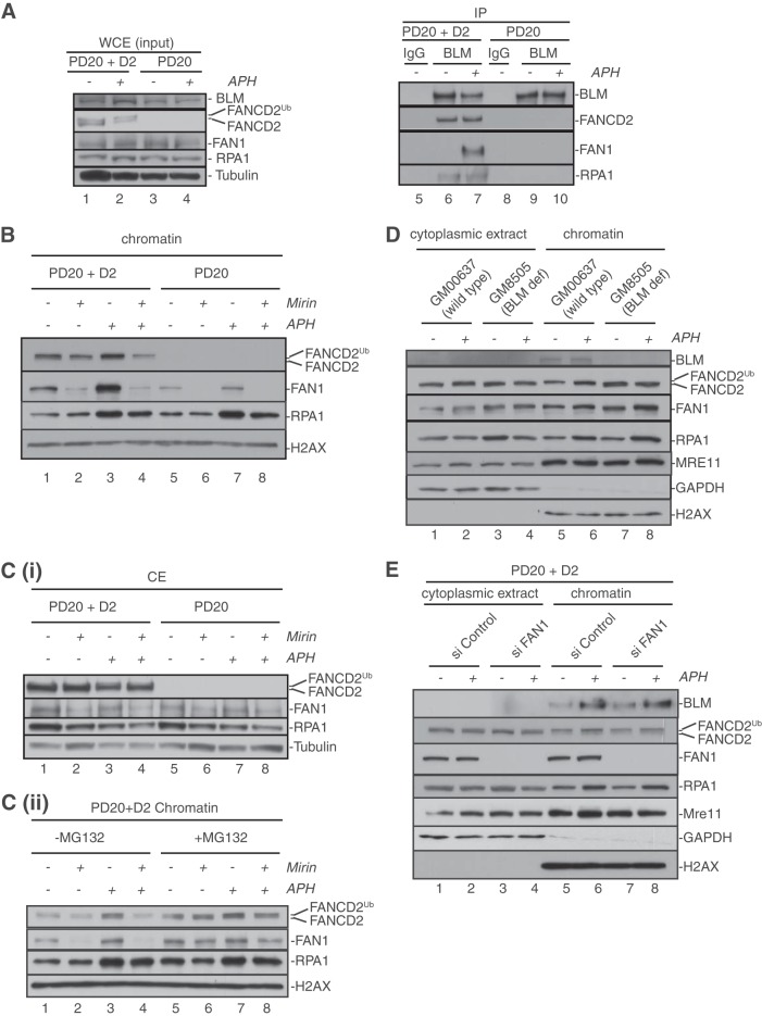
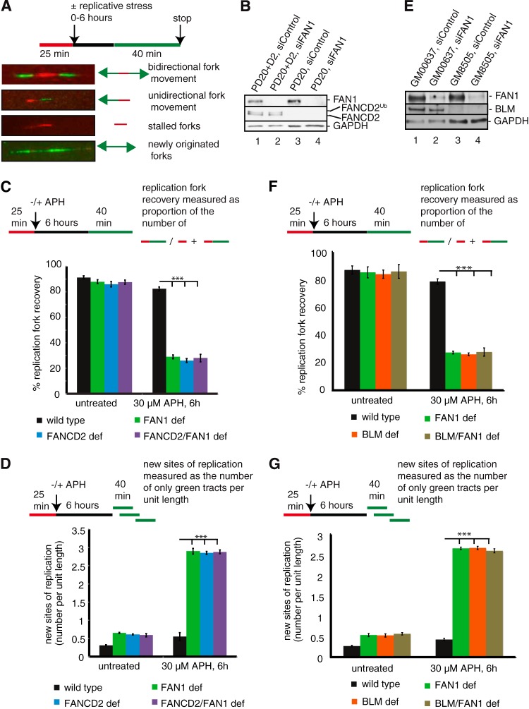
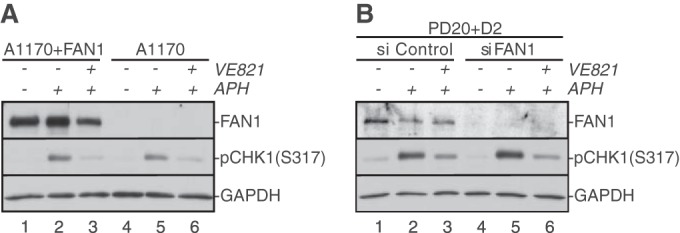
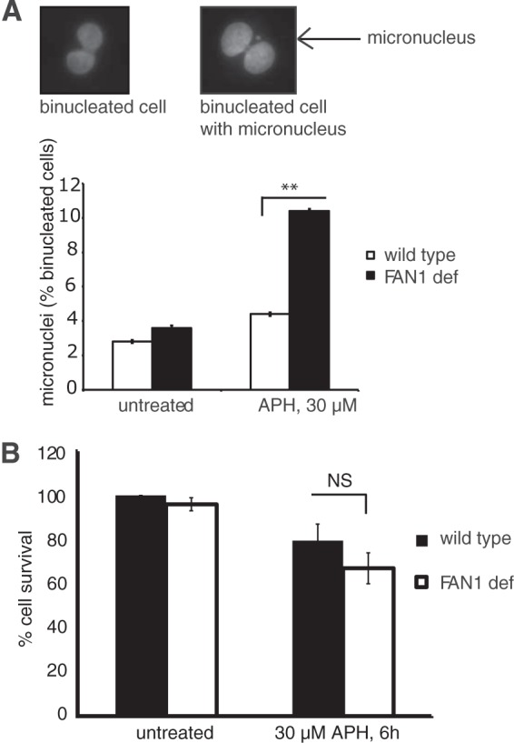
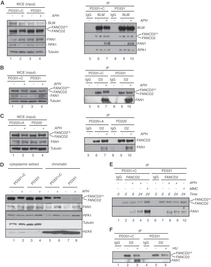
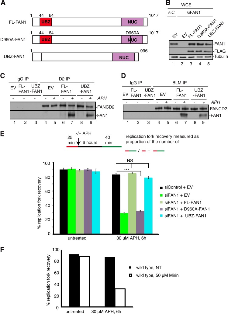
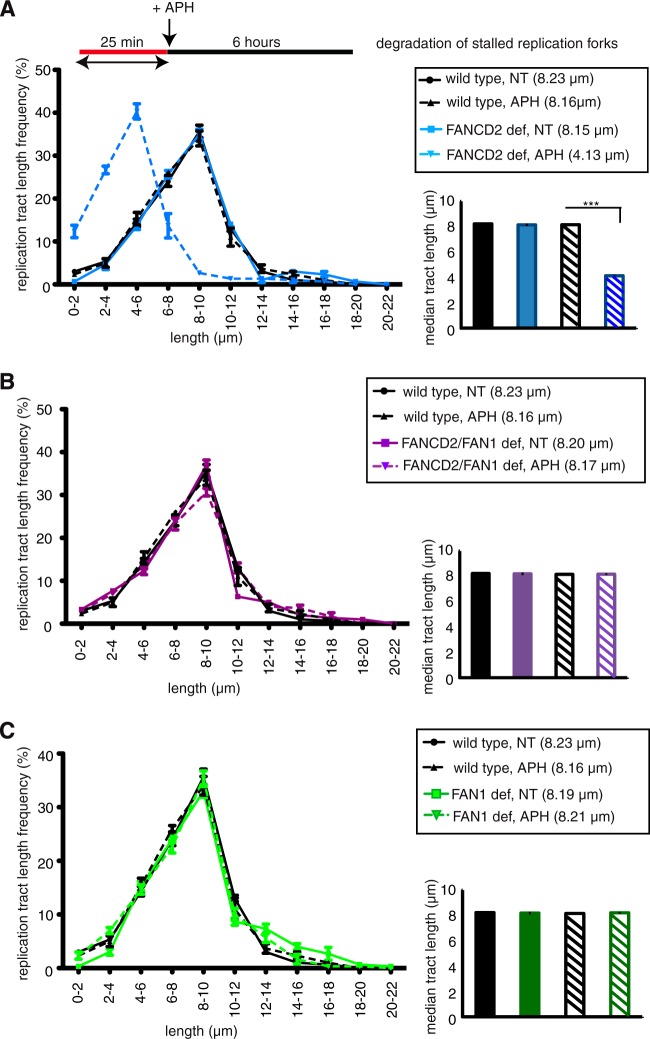
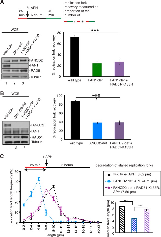
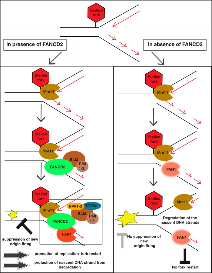
Fanconi anemia (FA) is a cancer predisposition syndrome characterized by cellular hypersensitivity to DNA interstrand cross-links (ICLs). Within the FA pathway, an upstream core complex monoubiquitinates and recruits the FANCD2 protein to ICLs on chromatin. Ensuing DNA repair involves the Fanconi-associated nuclease 1 (FAN1), which interacts selectively with monoubiquitinated FANCD2 (FANCD2(Ub)) at ICLs. Importantly, FANCD2 has additional independent functions: it binds chromatin and coordinates the restart of aphidicolin (APH)-stalled replication forks in concert with the BLM helicase, while protecting forks from nucleolytic degradation by MRE11. We identified FAN1 as a new crucial replication fork recovery factor. FAN1 joins the BLM-FANCD2 complex following APH-mediated fork stalling in a manner dependent on MRE11 and FANCD2, followed by FAN1 nuclease-mediated fork restart. Surprisingly, APH-induced activation and chromatin recruitment of FAN1 occur independently of the FA core complex or the FAN1 UBZ domain, indicating that the FANCD2(Ub) isoform is dispensable for functional FANCD2-FAN1 cross talk during stalled fork recovery. In the absence of FANCD2, MRE11 exonuclease-promoted access of FAN1 to stalled forks results in severe FAN1-mediated nucleolytic degradation of nascent DNA strands. Thus, FAN1 nuclease activity at stalled replication forks requires tight regulation: too little inhibits fork restart, whereas too much causes fork degradation.


Figures

FIG 1
FIG 1
Replication fork stalling stimulates formation of a BLM-FANCD2-FAN1 complex and triggers FAN1 binding to chromatin in an MRE11- and FANCD2-dependent manner. (A) APH treatment induces formation of a BLM-FANCD2-FAN1 complex in a FANCD2-dependent manner. FANCD2-proficient cells (PD20+D2) (lanes 1 and 2) and FANCD2-deficient cells (PD20) (lanes 3 and 4) were either left untreated (lanes 1 and 3) or treated with 30 μM APH for 6 h (lanes 2 and 4). Whole-cell extracts from these cells (lanes 1 to 4) were further subjected to immunoprecipitation (IP) with rabbit IgG (lanes 5 and 8; negative control) or an anti-BLM antibody (lanes 6, 7, 9, and 10). WCEs and IP samples were analyzed by Western blot (WB) detection using antibodies against BLM, FANCD2, FAN1, and RPA1. (B) Recruitment and stabilization of FAN1 at APH-stalled forks depend on MRE11 exonuclease activity and FANCD2. FANCD2-proficient (PD20+D2) and -deficient (PD20) cells were either left untreated or incubated with the MRE11 exonuclease inhibitor mirin; additionally, cells were left untreated or treated with 30 μM APH for 6 h, as indicated. Chromatin fractions were isolated from the cells and analyzed by WB for the presence of FANCD2, FAN1, and RPA1. Histone H2AX was used as a loading control. (C) Inhibition of MRE11 exonuclease activity triggers proteasomal degradation of FAN1. (i) Cellular mirin treatment reduces FAN1 protein levels. Cytoplasmic extracts isolated simultaneously with the chromatin fractions shown in panel B were analyzed by WB for the presence of FANCD2, FAN1, and RPA1. Tubulin was used as a loading control. (ii) Cellular treatment with MG132 restores FAN1 chromatin binding. FANCD2-proficient (PD20+D2) cells were left untreated or incubated with mirin; additionally, cells were left untreated or treated with 30 μM APH for 6 h, as indicated. Three hours after the addition of APH, cells were additionally either left untreated or treated with 10 μM MG132. Chromatin fractions were isolated from the cells and analyzed by WB for the presence of FANCD2, FAN1, and RPA1. Histone H2AX was used as a loading control. (D) Recruitment of FAN1 to APH-stalled forks occurs independently of BLM. BLM-proficient (GM00637) and BLM-deficient (GM08505) cells were left untreated or treated with 30 μM APH for 6 h, as indicated. Cytoplasmic and chromatin fractions were isolated from the cells and analyzed by WB for the presence of BLM, FANCD2, FAN1, and MRE11. GAPDH and histone H2AX were used loading controls for the cytoplasmic and chromatin fractions, respectively. (E) Recruitment of BLM to APH-stalled forks occurs independently of FAN1. FAN1-proficient and -deficient cells were left untreated or treated with 30 μM APH for 6 h, as indicated. Cytoplasmic and chromatin fractions were isolated and analyzed by WB for the presence of BLM, FANCD2, FAN1, and MRE11. GAPDH and histone H2AX were used as loading controls for the cytoplasmic and chromatin fractions, respectively. (Note that RPA1 was used in panels B to E as a positive control for the cellular response to APH.)
FIG 2
FIG 2
FAN1 acts in the same pathway with FANCD2 and BLM to mediate restart of APH-stalled replication forks and suppression of new origin firing. (A) Schematic of DNA fibers depicting sites of replication. Red tracts, DigU; green tracts, BioU. (B) Cell types used for DNA fiber analysis in panels C and D. Cells included wild-type (PD20+D2, siControl), FAN1-deficient (PD20+D2, siFAN1), FANCD2-deficient (PD20, siControl), and FANCD2-FAN1-double-deficient (PD20, siFAN1) cells. (C) FAN1 and FANCD2 act in a common pathway to mediate replication fork restart after APH-induced fork blockade. The efficiencies of replication restart in wild-type, FAN1-deficient, FANCD2-deficient, and FANCD2-FAN1-double-deficient cells were measured as the number of restarted replication forks (DigU-BioU tracts) compared with the total number of DigU-labeled tracts (DigU plus DigU-BioU). (D) FAN1 and FANCD2 act in concert to suppress new origin firing during replication blockade. The numbers of new sites of replication originating during the 40-min recovery period after APH treatment were compared between wild-type, FAN1-deficient, FANCD2-deficient, and FANCD2-FAN1-double-deficient cells. New origins of replication were measured as the number of green-only (BioU) tracts per unit length. (E) Cell types used for DNA fiber analysis in panels F and G. Cells included wild-type (GM00637, siControl), FAN1-deficient (GM00637, siFAN1), BLM-deficient (GM08505, siControl), and BLM-FAN1-double-deficient (GM08505, siFAN1) cells. (F) FAN1 and BLM act in a common pathway to mediate replication fork restart after APH-induced fork blockade. The efficiencies of replication restart in wild-type, BLM-deficient, FAN1-deficient, and BLM-FAN1-double-deficient cells were measured as described for panel C. (G) FAN1 and BLM act in concert to suppress new origin firing during replication blockade. The numbers of new sites of replication originating during the 40-min recovery period after APH treatment were compared between wild-type, BLM-deficient, FAN1-deficient, and BLM-FAN1-double-deficient cells. New origins of replication were measured as the number of green-only (BioU) tracts per unit length.
FIG 3
FIG 3
FAN1 does not regulate ATR activation in response to APH-triggered replication fork stalling. (A) Patient-derived FAN1-proficient (A1170+FAN1) or -deficient (A1170) cells were left untreated (lanes 1 and 4) or treated with 30 μM APH for 6 h (lanes 2, 3, 5, and 6). Additionally, cells were treated with dimethyl sulfoxide (DMSO) (lanes 2 and 5) or with the ATR inhibitor VE821 (lanes 3 and 6) 1 h prior to addition of APH. (B) Wild-type (PD20+D2, siControl) and FAN1-deficient (PD20+D2, siFAN1) cells were left untreated (lanes 1 and 4) or treated with 30 μM APH for 6 h (lanes 2, 3, 5, and 6) in the absence (lanes 2 and 5) or presence (lanes 3 and 6) of VE821. In panels A and B, WCE were analyzed for the presence of FAN1 and pCHK1-S317. GAPDH was used as a loading control.
FIG 4
FIG 4
FAN1 prevents APH-induced MN formation. (A) FAN1-deficient cells show increased MN formation in response to APH. Wild-type (PD20+D2, siControl) and FAN1-deficient (PD20+D2, siFAN1) cells were either left untreated or treated with 30 μM APH for 6 h, followed by a recovery period of 16 h. Binucleated G1-phase cells were counted for the presence of MN by using the cytokinesis block micronucleus assay (23). (Top) Representative images of a binucleated cell without an MN (left) and with an MN (right). (Bottom) Average MN frequencies are shown for wild-type and FAN1-deficient cells under unperturbed conditions and following APH treatment. (B) FAN1 deficiency does not reduce cell viability in response to APH. Wild-type (PD20+D2, siControl) and FAN1-deficient (PD20+D2, siFAN1) cells were left untreated or treated with 30 μM APH for 6 h. Cellular survival was measured by colony formation assay. Results were plotted as averages of results from two independent experiments.
FIG 5
FIG 5
FAN1 joins the BLM-FANCD2 complex at APH-stalled replication forks independently of FANCD2 monoubiquitination. (A) APH-triggered formation of the BLM-FANCD2-FAN1 complex occurs independently of a functional FA core complex. FANCC-proficient cells (PD331+C) (lanes 1 and 2) or FANCC-deficient cells (PD331) (lanes 3 and 4) were left untreated (lanes 1 and 3) or treated with 30 μM APH for 6 h (lanes 2 and 4). Whole-cell extracts from these cells (lanes 1 to 4) were further subjected to IP with rabbit IgG (lanes 5 and 8; negative control) or an anti-BLM antibody (lanes 6, 7, 9, and 10). WCEs and IP samples were analyzed by WB using antibodies against BLM, FANCD2, FAN1, and RPA1. (B and C) APH-triggered interaction of FAN1 with FANCD2 does not depend on the FA core complex members FANCC and FANCA. (B) FANCC-proficient cells (PD331+C) (lanes 1 and 2) or FANCC-deficient cells (PD331) (lanes 3 and 4) were either left untreated (lanes 1 and 3) or treated with 30 μM APH for 6 h (lanes 2 and 4). Whole-cell extracts from these cells (lanes 1 to 4) were further subjected to IP with rabbit IgG (lanes 5 and 8; negative control) or an anti-FANCD2 antibody (lanes 6, 7, 9, and 10). WCEs and IP samples were analyzed by WB using antibodies against FANCD2 and FAN1. (C) The same IP experiment as that described for panel B was carried out in FANCA-proficient (PD220+A) versus FANCA-deficient (PD220) cells. (D) Recruitment of FAN1 to APH-stalled forks occurs independently of FANCD2 monoubiquitination. FANCC-proficient (PD331+C) and -deficient (PD331) cells were left untreated or treated with 30 μM APH for 6 h, as indicated. Cytoplasmic and chromatin fractions were isolated and analyzed by WB for the presence of FANCD2 and FAN1. RPA1 was used as a positive control for the cellular response to APH. Tubulin and histone H2AX were used as loading controls for the cytoplasmic and chromatin fractions, respectively. (E) The MMC- but not APH-triggered FANCD2-FAN1 interaction depends on a functional FA core complex. FANCC-proficient (PD331+C) (lanes 1 to 5) or -deficient (PD331) (lanes 6 to 10) cells were left untreated, treated with 1 μM MMC, or treated with 30 μM APH, as indicated. Cell extracts were subjected to IP with mouse IgG (lanes 1 and 6; negative control) or an anti-FANCD2 antibody (lanes 2 to 5 and 7 to 10). IP samples were analyzed for the presence of FANCD2 and FAN1. (F) The HU-triggered FANCD2-FAN1 interaction depends on a functional FA core complex. FANCC-proficient cells (PD331+C) (lanes 1 to 3) or FANCC-deficient cells (PD331) (lanes 4 to 6) were either left untreated (lanes 1, 2, 4, and 5) or treated with 2 mM HU (lanes 3 and 6) for 24 h. WCEs from these cells were subjected to IP with mouse IgG (lanes 1 and 4; negative control) or an anti-FANCD2 antibody (lanes 2, 3, 5, and 6). IP samples were analyzed for the presence of FANCD2 and FAN1.
FIG 6
FIG 6
Functional cross talk between FAN1 and the BLM-FANCD2 complex during replication fork recovery requires FAN1 nuclease activity but not the FAN1 UBZ domain. (A) Schematic of wild-type and mutant FAN1 proteins used in the replication restart experiments. FL-FAN1, full-length wild-type FAN1; D960A-FAN1, full-length nuclease-dead FAN1 carrying a single amino acid substitution (D960A) in the nuclease domain (NUC); ΔUBZ-FAN1, truncated FAN1 lacking the N-terminal UBZ domain (21 amino acids). (B) Cell types used for DNA fiber analysis. Wild-type (PD20+D2) cells were stably transfected with either empty vector (EV) or a plasmid encoding siRNA-resistant, FLAG-tagged FL-FAN1, D960A-FAN1, or ΔUBZ-FAN1. The cells were then treated with control siRNA (siC) or FAN1 siRNA (siFAN1) for 72 h. WCEs were analyzed by WB for the presence of endogenous FAN1 (anti-FAN1 antibody) or FLAG-tagged FAN1 (anti-FLAG antibody). Tubulin was used as a loading control. (C) APH-triggered interaction of FAN1 with FANCD2 occurs independently of the FAN1 UBZ domain. Wild-type cells (PD20+D2) were transfected with either empty vector (EV; negative control) or a vector encoding siRNA-resistant, full-length FAN1 (FL-FAN1) or a FAN1 mutant lacking the UBZ domain (ΔUBZ-FAN1), followed by siFAN1 treatment to deplete endogenous FAN1 protein levels. Cells were either left untreated (lanes 1 to 4, 6, and 8) or treated with 30 μM APH for 6 h (lanes 5, 7, and 9) and then subjected to IP with rabbit IgG (lanes 1 to 3; negative control) or an anti-FANCD2 antibody (lanes 4 to 9). IP samples were analyzed by WB for the presence of FANCD2 and FAN1. (D) APH-triggered formation of the BLM-FANCD2-FAN1 complex occurs independently of the FAN1 UBZ domain. Wild-type cells were prepared as described for panel C and then subjected to IP with rabbit IgG (lanes 1 to 3; negative control) or an anti-BLM antibody (lanes 4 to 9). IP samples were analyzed by WB for the presence of FANCD2 and FAN1. (E) Analysis of replication fork restart following APH-triggered replication blockade in the cell types analyzed in panel B. The efficiencies of replication fork restart were compared between wild-type cells (siControl + EV), FAN1-deficient cells (siFAN1 + EV), and cells expressing exclusively FL-FAN1, D960A-FAN1, or ΔUBZ-FAN1. Fork restart efficiency was measured as the number of restarted replication forks (DigU-BioU tracts) compared with the total number of DigU-labeled tracts (DigU plus DigU-BioU tracts). (F) Mre11 exonuclease activity is required to mediate restart of APH-stalled replication forks. The efficiency of replication fork restart following APH-induced fork stalling was analyzed in wild-type (PD20+D2) cells in the presence or absence (NT) of the MRE11 exonuclease inhibitor mirin (50 μM). Restart efficiency was measured as the number of restarted replication forks (DigU-BioU tracts) compared with the total number of DigU-labeled tracts (DigU plus DigU-BioU tracts).
FIG 7
FIG 7
FAN1 degrades nascent DNA strands at APH-stalled replication forks in the absence of FANCD2. Lengths of nascent replication fork tracts indicating fork stability (labeled with DigU only) were measured before (NT) and after 6 h of APH treatment. Preformed DigU tract lengths shortened during APH treatment in FANCD2-deficient (PD20) cells compared to wild-type (PD20+D2) cells (A) but not in FANCD2-FAN1-double-deficient (PD20, siFAN1) cells compared to wild-type (PD20+D2) cells (B). (C) Preformed DigU tract lengths do not shorten in FAN1-deficient (PD20+D2, siFAN1) cells. (Insets) Plotted median tract lengths.
FIG 8
FIG 8
RAD51-K133R overexpression does not compensate for replication restart defects in FAN1- or FANCD2-deficient cells. (A) RAD51-K133R does not rescue replication fork restart in FAN1-deficient cells. The efficiencies of replication restart in wild-type cells (A1170+FAN1) (lane 1), FAN1-deficient cells (A1170) (lane 2), and FAN1-deficient cells expressing RAD51-K133R (A1170+RAD51-K133R) (lane 3) were measured as the number of restarted replication forks (DigU-BioU tracts) compared with the total number of DigU-labeled tracts (DigU plus DigU-BioU tracts). (B) RAD51-K133R does not rescue replication fork restart in FANCD2-deficient cells. The efficiencies of replication restart in wild-type cells (PD20+D2) (lane 1), FANCD2-deficient cells (PD20) (lane 2), and FANCD2-deficient cells expressing RAD51-K133R (PD20+RAD51-K133R) (lane 3) were measured as the number of restarted replication forks (DigU-BioU tracts) compared with the total number of DigU-labeled tracts (DigU plus DigU-BioU tracts). (C) RAD51-K133R promotes replication fork stability in FANCD2-deficient cells. Lengths of nascent replication fork tracts indicating fork stability (labeled with DigU only) were measured after 6 h of APH treatment. Preformed DigU tract lengths shortened during APH treatment in FANCD2-deficient cells (median length = 4.71 μm) compared to wild-type cells (median length = 8.62 μm). DigU tract length shortening was counteracted by expression of RAD51-K133R in FANCD2-deficient cells (median length = 7.56 μm).
FIG 9
FIG 9
Model describing the role of FAN1 at a stalled replication fork. (A) Role of FAN1 in the presence of FANCD2. MRE11 is recruited to APH-stalled replication forks first, followed by FANCD2 and its constitutive interaction partner, BLM. Nonubiquitinated FANCD2 and MRE11 then both support the recruitment of FAN1. Subsequently, all four proteins (likely in concert with other BLM complex members) act in concert to promote replication fork restart in a manner dependent on the MRE11 and FAN1 nuclease activities. Concurrent with efficient fork restart, the firing of new origins is suppressed. In addition, FANCD2 protects the nascent DNA strands from nucleolytic attack by MRE11 and FAN1 during replication fork stalling. (B) Role of FAN1 in the absence of FANCD2. MRE11 is recruited to APH-stalled forks and enables partial recruitment of FAN1 independently of FANCD2. Despite the presence of MRE11 and FAN1, replication forks cannot restart efficiently, which in turn triggers firing of new replication origins. At the same time, the absence of FANCD2 allows for uncontrolled access of MRE11 and FAN1 to DNA at the stalled fork, leading to nucleolytic degradation of nascent DNA strands behind the fork.

References

    1. Kee Y, D'Andrea AD. 2012. Molecular pathogenesis and clinical management of Fanconi anemia. J. Clin. Invest. 122:3799–3806. 10.1172/JCI58321. - DOI - PMC - PubMed
    1. Kupfer GM. 2013. Fanconi anemia: a signal transduction and DNA repair pathway. Yale J. Biol. Med. 86:491–497. - PMC - PubMed
    1. Naim V, Rosselli F. 2009. The FANC pathway and BLM collaborate during mitosis to prevent micro-nucleation and chromosome abnormalities. Nat. Cell Biol. 11:761–768. 10.1038/ncb1883. - DOI - PubMed
    1. Kee Y, D'Andrea AD. 2010. Expanded roles of the Fanconi anemia pathway in preserving genomic stability. Genes Dev. 24:1680–1694. 10.1101/gad.1955310. - DOI - PMC - PubMed
    1. Schlacher K, Wu H, Jasin M. 2012. A distinct replication fork protection pathway connects Fanconi anemia tumor suppressors to RAD51-BRCA1/2. Cancer Cell 22:106–116. 10.1016/j.ccr.2012.05.015. - DOI - PMC - PubMed

Publication types

MeSH terms

LinkOut - more resources