

Transcorneal electrical stimulation in patients with retinal artery occlusion: a prospective, randomized, sham-controlled pilot study
- PMID: 25135699
- PMCID: PMC4108151
- DOI: 10.1007/s40123-013-0012-5
Transcorneal electrical stimulation in patients with retinal artery occlusion: a prospective, randomized, sham-controlled pilot study
Abstract
Introduction: The purpose of this study was to investigate the safety and efficacy of transcorneal electrical stimulation (TES) in patients suffering from retinal artery occlusion (RAO).
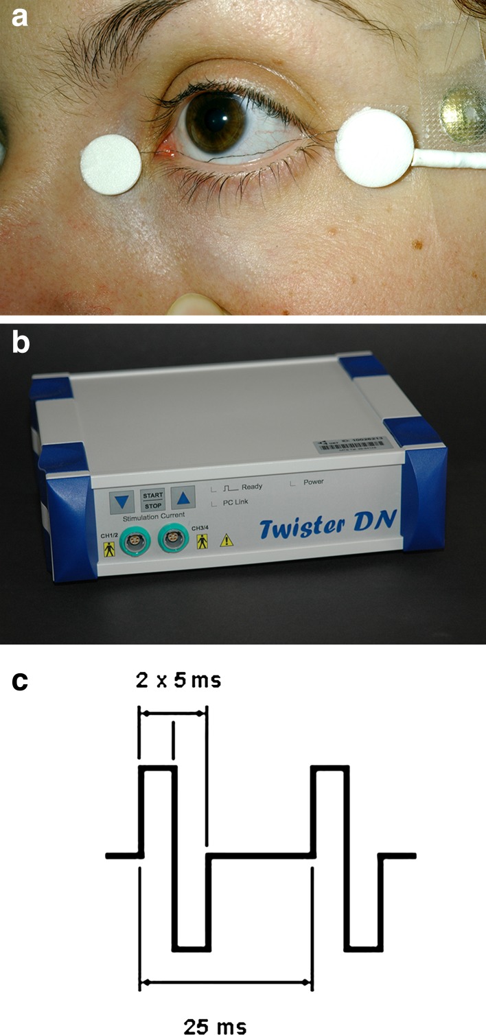
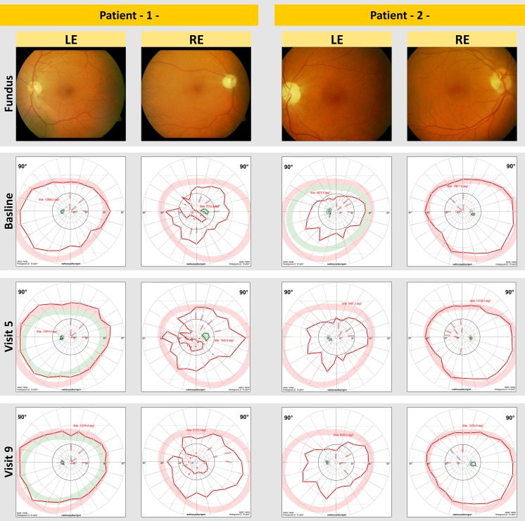
Methods: Twelve patients with central and one patient with branch RAO (age 25-84 years, median 74 years) were enrolled in this prospective, randomized, sham-controlled study. RAO was diagnosed 10 days to 17 months prior to study participation. Patients were treated with TES (5 ms positive followed by 5 ms negative biphasic pulses at 20 Hz; applied with DTL electrodes) for 30 min once a week for 6 consecutive weeks. Patients were randomly assigned to TES with 0 mA (sham, n = 3), 66% (n = 5) or 150% (n = 5) of the patient's individual electrical phosphene threshold (EPT) at 20 Hz. Best corrected visual acuity, ophthalmology examination and EPT (at 3, 6, 9, 20, 40, 60, and 80 Hz) were determined at baseline and at eight follow-up visits over 17 weeks. During four visits (week 1, 5, 9, and 17) kinetic and static visual fields as well as full-field and multifocal electroretinography were measured. The method of restricted maximum likelihood (P < 0.05, Tukey-Kramer) was used to estimate the development of parameters under treatment.
Results: TES was tolerated well; no ocular or systemic adverse events were observed except for foreign-body sensation after TES (n = 3). During the study period the slopes of the scotopic a-wave increased significantly (high-intensity flash white 10 cd.s/m(2); P = 0.03) in the 150% treatment group. All other parameters in all other groups remained statistically unchanged.
Conclusions: Although TES was tolerated well, statistically significant improvements were found only for specific a-wave slopes. This is in contradiction to previous smaller, uncontrolled reports. Further studies with larger sample sizes and longer duration might, however, show additional significant effects.
Figures
References
-
- Von Graefe A. Über Embolie der Arteria centralis retinae als Ursache plötzlicher Erblindung. Graefes Arch Clin Exp Ophthalmol. 1859;5:136–185. doi: 10.1007/BF02720764. - DOI
LinkOut - more resources
Full Text Sources
Other Literature Sources
