. 2013 Jun;2(1):41-51.
doi: 10.1007/s40123-013-0014-3. Epub 2013 Jun 1.

Vascular endothelial growth factor in anterior chamber liquid patients with diabetic retinopathy, cataract and neovascular glaucoma

Affiliations

Vascular endothelial growth factor in anterior chamber liquid patients with diabetic retinopathy, cataract and neovascular glaucoma

Anatoly Kuzmin et al. Ophthalmol Ther. 2013 Jun.

Abstract

Introduction: The aims of this study were: (1) to investigate the association of vascular endothelial growth factor isoform A (VEGF-A) concentration in the anterior chamber liquid (ACL) with vascular proliferation in patients with diabetic retinopathy (DR) who had undergone surgical treatment for cataract and neovascular glaucoma; (2) to analyze the association of VEGF-A level in ACL with the cataract surgery outcomes.

Materials and methods: Undiluted aqueous fluid samples were obtained from 207 eyes of patients who underwent intraocular surgery, 136 patients with diabetes mellitus (DM) and 22 patients without DM. The ACL samples were obtained during operation. The VEGF-A levels were analyzed by enzyme-linked immunosorbent assay.

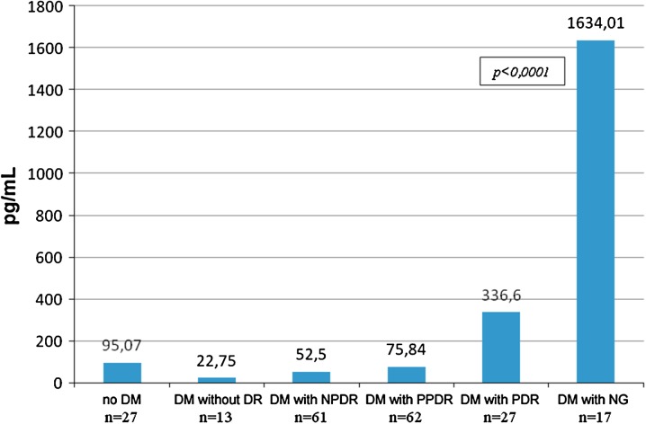
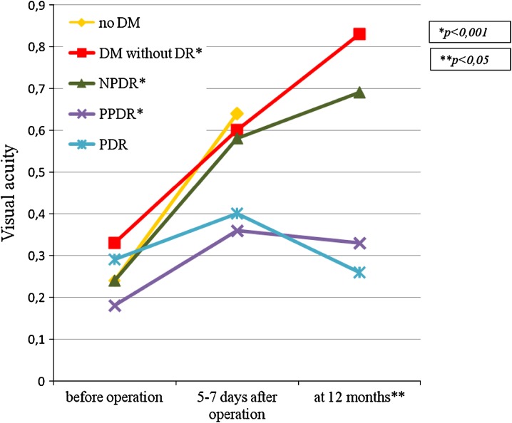
Results: The lowest VEGF-A levels were in diabetic patients without signs of DR [22.75 pg/mL (10.78; 63.36)]. More severe DR tended to occur in diabetic patients with higher VEGF-A levels in ACL. In diabetic patients with proliferative DR (PDR), VEGF-A levels were significantly higher [336.6 pg/mL (232.3; 410.74)] than in patients without DR P < 0.0001. In patients with terminal stage of DR [neovascular glaucoma (NG)], VEGF-A levels were dramatically higher and attained 1,634.01 pg/mL (610.69; 2657.33). In non-diabetic patients, VEGF-A levels were 95.07 pg/ml (60.92; 129.22). The best visual acuity results in post-operative period were observed in the group of diabetic patients without DR. In the group of patients with PDR, post-operative visual acuity [0.26 (0.1; 0.42)] was similar to visual acuity before operation [0.29 (0.13; 0.44)]. There was no significant increase in visual acuity due to cataract surgery. In 52.4% patients, no complications had occurred by the end of the follow-up period. In 40% patients, retinal laser coagulation was performed, and in 7.6% patients NG had developed.

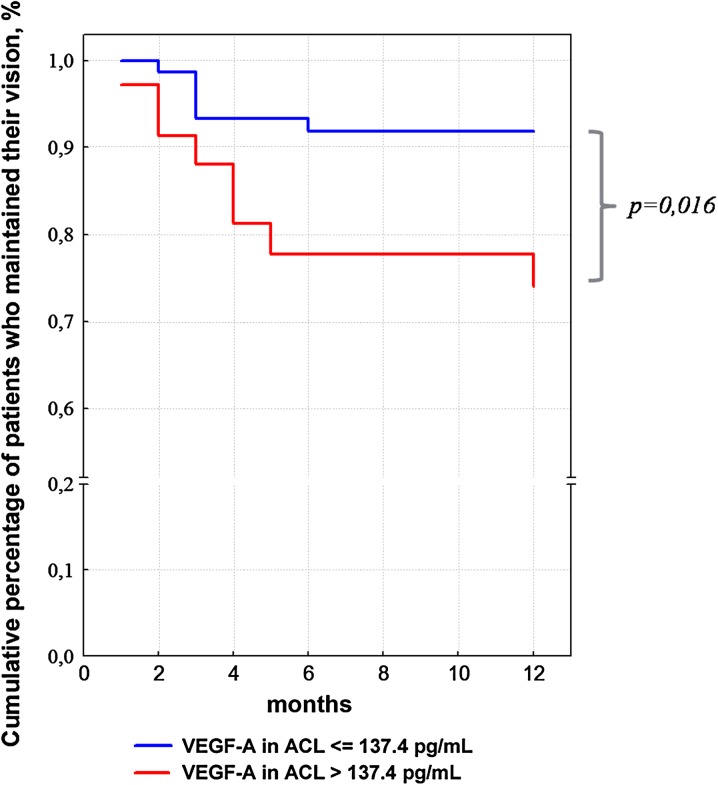
Conclusion: VEGF-A level in ACL increases with DR progression and may be of prognostic value in evaluating the potential risk of further neovascularization progression in diabetic patients.


Figures

Fig. 1
Fig. 1
Distribution of operated diabetic patients with cataract according to DR type [ examinations have been performed before (a) and after surgery (b)]. No DR no signs of diabetic retinopathy were detected, NPDR non-proliferative diabetic retinopathy, PPDR pre-proliferative diabetic retinopathy, PDR proliferative diabetic retinopathy
Fig. 2
Fig. 2
Median of VEGF-A levels in ACL in operated patients (pg/mL) as function of DR severity. Axis Y, VEGF-A in ACL. ACL anterior chamber liquid, DM diabetes mellitus, DR diabetic retinopathy, NPDR non-proliferative diabetic retinopathy, PPDR pre-proliferative diabetic retinopathy, PDR proliferative diabetic retinopathy, NG neovascular glaucoma, VEGF-A vascular endothelial growth factor A
Fig. 3
Fig. 3
Visual acuity changes in patients operated for cataract. DM diabetes mellitus, DR diabetic retinopathy, NPDR non-proliferative diabetic retinopathy, PPDR pre-proliferative diabetic retinopathy, PDR proliferative diabetic retinopathy, NG neovascular glaucoma. *P < 0.001—the significance of the differences between baseline visual acuity and visual acuity at 12 months after surgery (Wilcoxon test). **P < 0.05—the significance of the differences of the visual acuity at 12 months between groups (Mann–Whitney test with the Bonferroni adjustment). The authors checked the best-corrected visual acuity by the Golovin–Sivtsev table from a 5-m distance. It had been estimated in conventional units. For example, visual acuity 1.0 (Golovin–Sivtsev table) is equivalent to 20/20 (Snellen chart); visual acuity 0.1 (Golovin–Sivtsev table) is equivalent to 20/200 (Snellen chart)
Fig. 4
Fig. 4
Percentages of diabetic patients with different VEGF-A ACL levels in whom visual acuity at 12 months were maintained at the same level as post-operation. ACL anterior chamber liquid, VEGF-A vascular endothelial growth factor A

References

    1. World Health Organization. Sight test and glasses could dramatically improve the lives of 150 million people with poor vision. Press release, 11 October 2006. http://www.who.int/mediacentre/news/releases/2006/pr55/en/index.html (Accessed 1 Dec 2012). - PubMed
    1. Dagher Z, Park YS, Asnaghi V, et al. Studies of rat and human retinas predict a role for the polyol pathway in human diabetic retinopathy. Diabetes. 2004;53:2404–2411. doi: 10.2337/diabetes.53.9.2404. - DOI - PubMed
    1. Pournaras CJ, Rungger-Brändle E, Riva CE, Hardarson SH, Stefansson E. Regulation of retinal blood flow in health and disease. Prog Retin Eye Res. 2008;27:284–330. doi: 10.1016/j.preteyeres.2008.02.002. - DOI - PubMed
    1. The diabetes control and complications trial research group The affect of intensive treatment of diabetes on the development and progression of long-term complications in insulin-dependent diabetes mellitus. N Engl J Med. 1993;329:977–986. doi: 10.1056/NEJM199309303291401. - DOI - PubMed
    1. Aiello LP, Northrup JM, Keyt BA, Takagi H, Iwamoto MA. Hypoxic regulation of vascular endothelial growth factor in retinal cells. Arch Ophthalmol. 1995;113:1538–1544. doi: 10.1001/archopht.1995.01100120068012. - DOI - PubMed

LinkOut - more resources