. 2014 Oct 28:192:310-6.
doi: 10.1016/j.jconrel.2014.08.006. Epub 2014 Aug 15.

Micro-fractional epidermal powder delivery for improved skin vaccination

Affiliations

Micro-fractional epidermal powder delivery for improved skin vaccination

Xinyuan Chen et al. J Control Release. .

Abstract

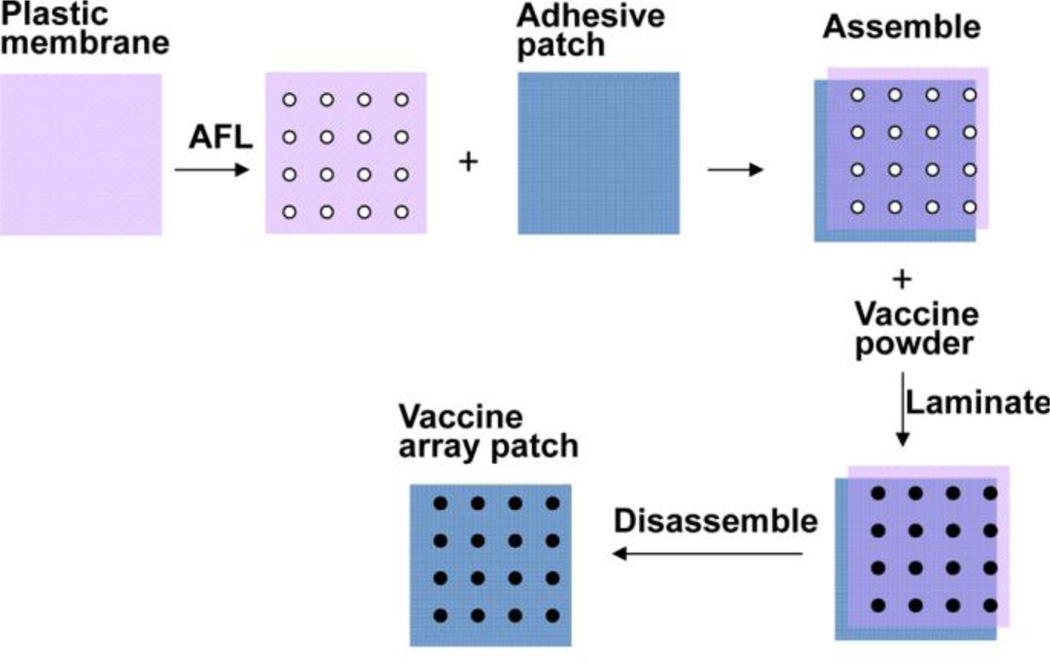
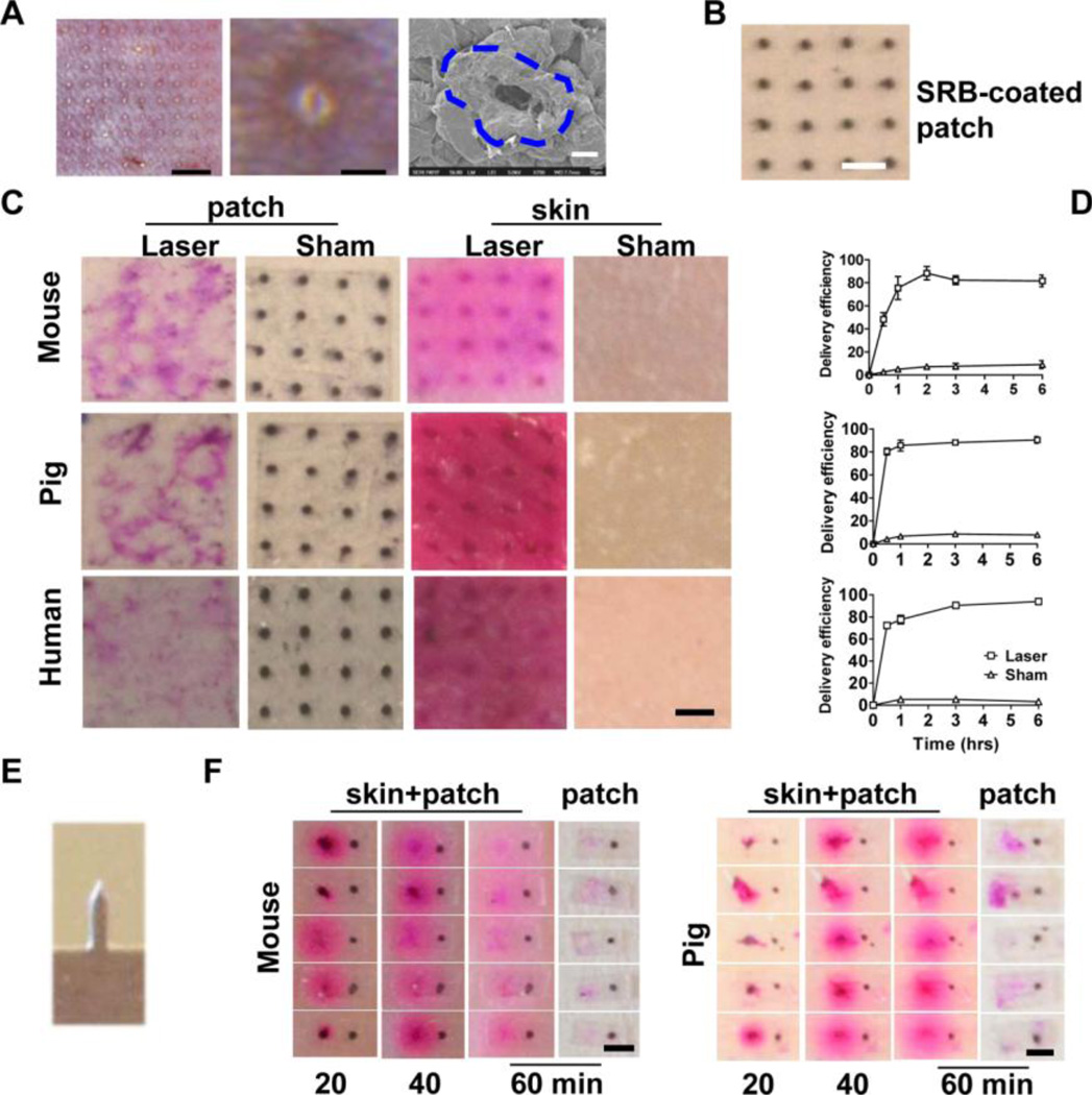
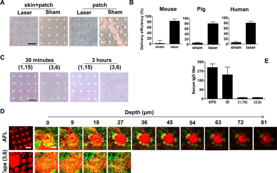
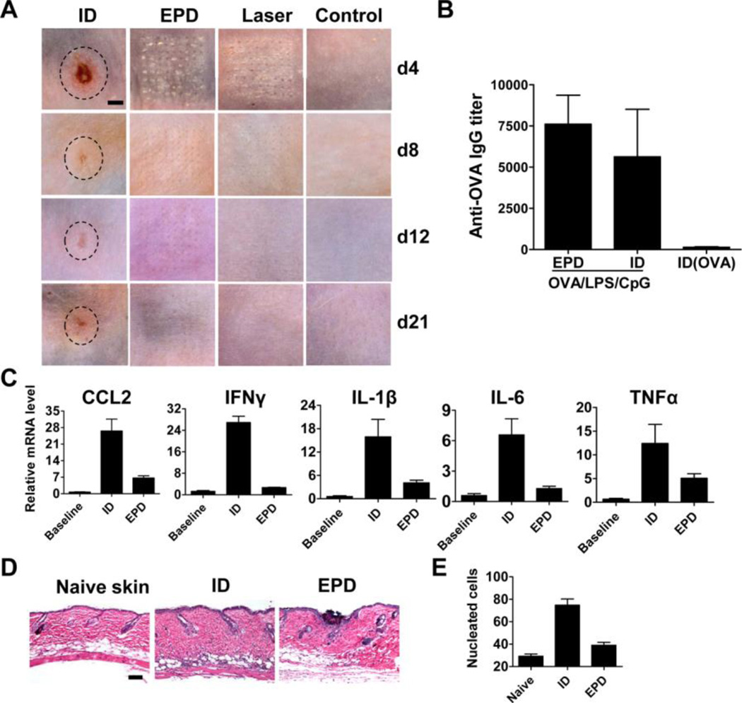
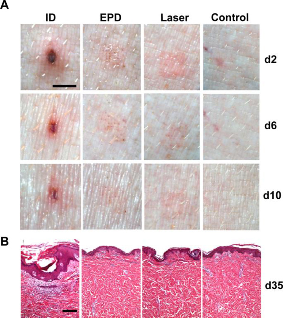
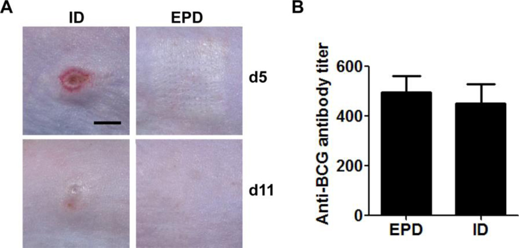
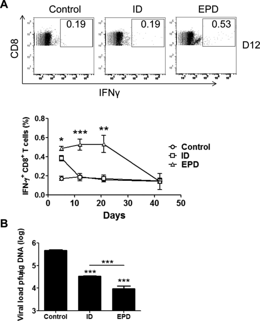
Skin vaccination has gained increasing attention in the last two decades due to its improved potency compared to intramuscular vaccination. Yet, the technical difficulty and frequent local reactions hamper its broad application in the clinic. In the current study, micro-fractional epidermal powder delivery (EPD) is developed to facilitate skin vaccination and minimize local adverse effects. EPD is based on ablative fractional laser or microneedle treatment of the skin to generate microchannel (MC) arrays in the epidermis followed by topical application of powder drug/vaccine-coated array patches to deliver drug/vaccine into the skin. The novel EPD delivered more than 80% sulforhodamine b (SRB) and model antigen ovalbumin (OVA) into murine, swine, and human skin within 1h. EPD of OVA induced anti-OVA antibody titer at a level comparable to intradermal (ID) injection and was much more efficient than tape stripping in both delivery efficiency and immune responses. Strikingly, the micro-fractional delivery significantly reduced local side effects of LPS/CpG adjuvant and BCG vaccine, leading to complete skin recovery. In contrast, ID injection induced severe local reactions that persisted for weeks. While reducing local reactogenicity, EPD of OVA/LPS/CpG and BCG vaccine generated a comparable humoral immune response to ID injection. EPD of vaccinia virus encoding OVA induced significantly higher and long-lasting interferon γ-secreting CD8+ T cells than ID injection. In conclusion, EPD represents a promising technology for needle-free, painless skin vaccination with reduced local reactogenicity and at least sustained immunogenicity.

Keywords: Laser; Local reactogenicity; Microneedle; Powder delivery; Skin vaccination; Vaccine adjuvant.


Conflict of interest statement

The authors have no conflict of interest to declare.

Figures

Figure 1
Figure 1. Illustration of powder array patch coating
A plastic membrane was exposed to laser illumination (35mJ, 5%) to generate 4×4 array of microholes in ~2×2 mm2 area, each with a measured diameter of 189µm. The membrane was topically layered onto an adhesive patch (3M). Vaccine powders were poured onto the membrane/patch assembly and pushed to fill the microholes. Non-adherent powders were removed before disassembly of the plastic membrane/adhesive patch assembly to obtain powder vaccine coated array patches.
Figure 2
Figure 2. EPD of SRB
A. Characteristics of laser-generated MCs in mouse skin. Left, laser generated a 9×9 MC array (scale: 1.5mm); Middle, enlarged protrusion with a tiny microhole at the center (scale: 125µm); Right, representative SEM image of a MC with elevated edges outlined by dashed line (scale: 20µm). B. Representative powder SRB-coated array patch. Scale: 750µm. C. SRB-coated patches were topically applied onto laser- or sham-treated mouse, pig, and human skin. Three hours later, patches were removed and representative patch and skin pictures were shown. Scale: 750µm. D. At indicated times, SRB-coated patches were removed and the delivery efficiency was calculated after quantification of SRB amount in the skin and on the patch. n=4. E. Image of one MN in a row of 5. F. A row of 5 MNs as in E. was used to pierce live mouse skin and dissected pig skin followed by topical application of 5 single row patches each with 2 SRB coating spots. One spot (on the left) was put just above MN-generated MC and the other (on the right) was put on intact skin surface. Images were taken 20, 40, and 60 minutes later. Patches were further removed and patch pictures were taken. Scale: 750µm.
Figure 3
Figure 3. EPD of OVA
A. Powder OVA-coated array patches were topically applied onto laser- or sham-treated skin. Three hours later, skin pictures with patch attached (skin+patch) and patch pictures (patch) after patch removal were shown. Scale: 750µm. B. OVA patches were removed 1 hour after application and the amount of OVA remained on the patches was measured to calculate the delivery efficiency. C. Skin was subjected to tape stripping followed by topical application of the same OVA array patches as in A. Thirty minutes and 3 hours later, skin pictures were taken. D. Powder AF647-OVA-coated array patches were topically applied onto laser-treated or tape (3, 6)-treated skin of MHC II-EGFP mice for 6 hours. Skin pictures were taken at superficial level (left panel, scale: 750µm) or at different depths (right panels, scale: 100µm) under intravital confocal microscope. Green: GFP-labeled APCs; red: AF647-OVA. E. Mice were immunized with EPD or tape stripping-based powder delivery of OVA or intradermally injected with the same amount of OVA. Two weeks later, serum anti-OVA IgG titer was measured. n=5.
Figure 4
Figure 4. EPD reduces local reactogenicity of LPS/CpG adjuvant in mice
A. Mice were treated with laser followed by topical application of LPS/CpG (20µg each)-coated array patches (EPD), or intradermally injected with patch extracts (ID), or treated with laser alone, or left untreated. Skin pictures were taken on day 4, 8, 12 and 21 and representative local reactions were shown. The edges of the local reactions in ID group were outlined by dashed lines. Scale: 1.5mm. B. Mice were treated with laser followed by topical application of OVA/LPS/CpG (10µg/20µg/20µg)-coated array patches (EPD) or intradermally injected with patch extracts (ID), or intradermally injected with the same amount of OVA (ID(OVA)). Two weeks later, serum anti-OVA IgG titer was measured. n=4. C. Mice were treated with laser followed by topical application of LPS/CpG-coated array patches (2µg each), or intradermally injected with the same amount of LPS/CpG mixture, or left untreated. 24 hours later, skin was dissected and expression of CCL2, IFNγ, IL-1β, IL-6 and TNFα was quantified. n=2–4. D. Mice were similarly treated as in C. 24 hours later, skin was dissected and subjected to histological analysis. Representative H&E-stained sections were shown. Scale: 100µm. E. Total nucleated cells in sections from D. were calculated. n=5–7.
Figure 5
Figure 5. EPD reduces local reactogenicity of LPS/CpG adjuvant in pigs
Pigs were treated with laser followed by topical application of LPS/CpG(20µg each)-coated array patches, or intradermally injected with LPS/CpG from patch extracts, or treated with laser alone, or intradermally injected with PBS. A. Representative local reactions on day 2, 6, and 10 were shown. Scale: 3mm. B. pigs were sacrificed on day 35 and skin samples were collected and subjected to histological analysis. Representative skin H&E sections were shown. Scale: 200µm.
Figure 6
Figure 6. EPD reduces local side effects of BCG vaccine in mice
Mice were treated with laser followed by topical application of BCG vaccine-coated array patches or intradermally injected with BCG vaccine from patch extracts. Skin reactions were monitored daily and representative local reactions on day 5 and 11 are shown in A (scale: 3mm). Serum anti-BCG antibody titer was measured 2 weeks after immunization and shown in B. n=4
Figure 7
Figure 7. EPD improves immunogenicity of VV-OVA in mice
A. Mice were immunized with EPD of VV-OVA or intradermally injected with the same amount of VV-OVA. 5, 12, 21 days later, percentage of IFNγ-secreting CD8+ T cells in PBMCs was analyzed by flow cytometry. Representative flow cytometry figures (upper) and the percentage of IFNγ-secreting CD8+ T cells (lower) were shown. B. Mice were challenged 6 week after immunization and viral load 6 days after challenge was shown. n=4.

References

    1. Kim YC, Prausnitz MR. Enabling skin vaccination using new delivery technologies. Drug Deliv. Transl. Res. 2011;1:7–12. - PMC - PubMed
    1. Chen D, Maa YF, Haynes JR. Needle-free epidermal powder immunization. Expert. Rev. Vaccines. 2002;1:265–276. - PubMed
    1. Glenn GM, Rao M, Matyas GR, Alving CR. Skin immunization made possible by cholera toxin. Nature. 1998;391:851. - PubMed
    1. Karande P, Mitragotri S. Transcutaneous immunization: an overview of advantages, disease targets, vaccines, and delivery technologies. Annu. Rev. Chem. Biomol. Eng. 2010;1:175–201. - PubMed
    1. Kendall MA. Needle-free vaccine injection. Handb. Exp. Pharmacol. 2010:193–219. - PubMed

Publication types