. 2014 Sep 15;193(6):3080-9.
doi: 10.4049/jimmunol.1303196. Epub 2014 Aug 18.

SARS-coronavirus open reading frame-9b suppresses innate immunity by targeting mitochondria and the MAVS/TRAF3/TRAF6 signalosome

Affiliations

SARS-coronavirus open reading frame-9b suppresses innate immunity by targeting mitochondria and the MAVS/TRAF3/TRAF6 signalosome

Chong-Shan Shi et al. J Immunol. .

Abstract

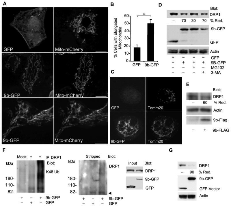
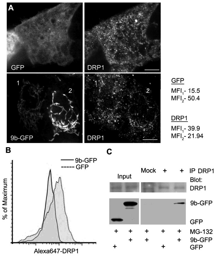
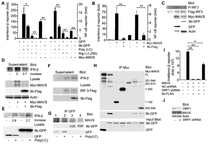
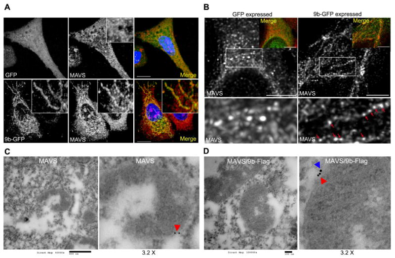
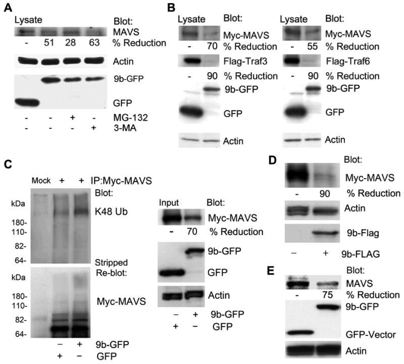
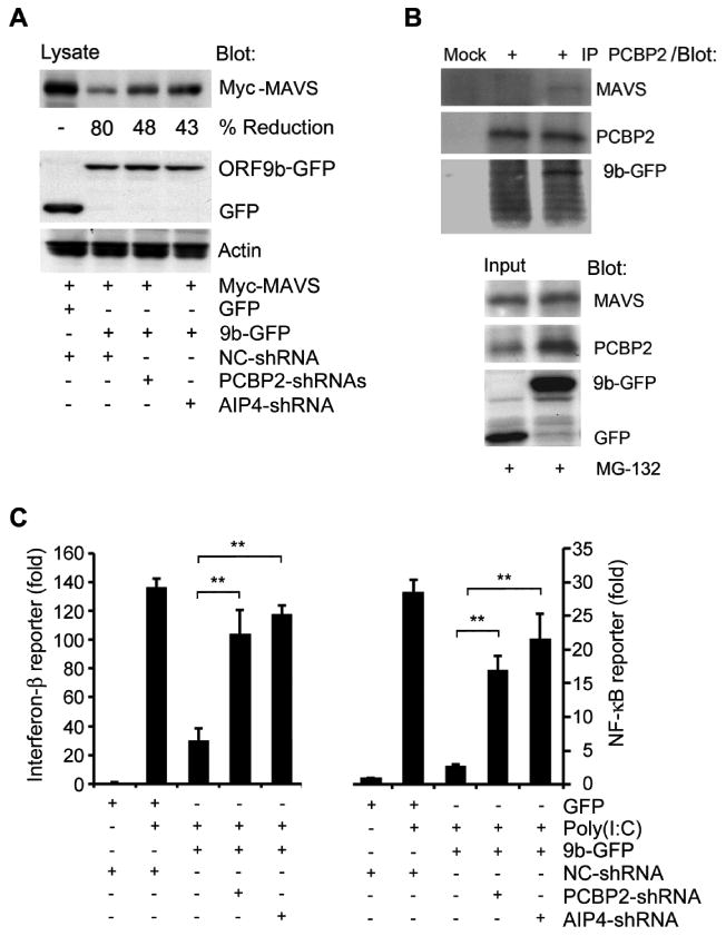
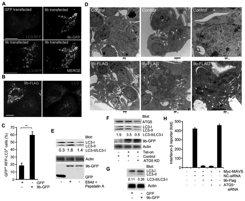
Coronaviruses (CoV) have recently emerged as potentially serious pathogens that can cause significant human morbidity and death. The severe acute respiratory syndrome (SARS)-CoV was identified as the etiologic agent of the 2002-2003 international SARS outbreak. Yet, how SARS evades innate immune responses to cause human disease remains poorly understood. In this study, we show that a protein encoded by SARS-CoV designated as open reading frame-9b (ORF-9b) localizes to mitochondria and causes mitochondrial elongation by triggering ubiquitination and proteasomal degradation of dynamin-like protein 1, a host protein involved in mitochondrial fission. Also, acting on mitochondria, ORF-9b targets the mitochondrial-associated adaptor molecule MAVS signalosome by usurping PCBP2 and the HECT domain E3 ligase AIP4 to trigger the degradation of MAVS, TRAF3, and TRAF 6. This severely limits host cell IFN responses. Reducing either PCBP2 or AIP4 expression substantially reversed the ORF-9b-mediated reduction of MAVS and the suppression of antiviral transcriptional responses. Finally, transient ORF-9b expression led to a strong induction of autophagy in cells. The induction of autophagy depended upon ATG5, a critical autophagy regulator, but the inhibition of MAVS signaling did not. These results indicate that SARS-CoV ORF-9b manipulates host cell mitochondria and mitochondrial function to help evade host innate immunity. This study has uncovered an important clue to the pathogenesis of SARS-CoV infection and illustrates the havoc that a small ORF can cause in cells.


Figures

Figure 1
Figure 1
ORF-9b expression elongates mitochondria and enhances the degradation of DRP1. (A) Confocal microscopy of A549 cells expressing GFP (top) or 9b-GFP (middle) and mito-mCherry or HEK 293 cells expressing 9b-GFP and mito-mCherry (lower). Individual images are shown. Scale bar: 10 μm. (B) Quantitation of percentage of cells with elongated mitochondria in A549 cells expressing GFP or 9b-GFP and Mito-mCherry. Co-expressing cells (100 each) were imaged and % of cells with obviously elongated mitochondria enumerated. (C) Confocal microscopy of THP-1 cells permanently expressing 9b-GFP or GFP and immunostained for Tomm20. (D) Immunoblot of cell lysates from HEK 293 cells expressing GFP or 9b-GFP to detect DRP1. In some instances the cells were treated with MG-132 (10 μM)) or 3-MA (5 mM) for 4 h. (E) Immunoblots of cell lysates from HEK 293 cells transfected with Flag or 9b-Flag for indicated proteins. (F) Immunoblots of DRP1 immunoprecipitates (+) and cell lysates from HEK 293 cells transfected with GFP or 9b-GFP. The mock lane is a control antibody immunoprecipitation. Following the K48 ubiquitin (Ub) immunoblot the membrane was stripped and re-blotted for DRP1. The cells were treated with MG-132 for the last 4 h prior to cell lysis. (G) Immunoblot of cell lysates from THP-1 cells permanently expressing either 9b-GFP or GFP to detect DRPl.
Figure 2
Figure 2
ORF-9b expression leads to less DRP1 associated with mitochondria and ORF-9b co-immunoprecipitates with DRP1. (A) Confocal microscopy of endogenous DRP1 in A549 cells transiently transfected with GFP-ORF9b or GFP vector alone. Scale bar: 10 μm. Mean fluorescence intensity (MFI) of DRP1 or GFP was quantified from the whole volume using ImageJ for cell 1 and cell 2. (B) Flow cytometry analysis of endogenous DRP1 level in A549 cells expressing either GFP or 9b-GFP. The mean fluorescent intensity of DRP1 in GFP+ cells was 186 and 46 in the 9b-GFP+ cells. (C) Immunoblots of DRP1 immunoprecipitates (+) and cell lysates from HEK 293 cells transfected with GFP or 9b-GFP for indicated proteins. Mock is a control antibody immunoprecipitation. Cells were treated with MG-132 (10 μM).
Figure 3
Figure 3
ORF-9b inhibits anti-viral type I interferon responses. (A) IFN-β and NF-κB reporter gene assays using HEK 293 cells expressing GFP or 9b-GFP and induced by transfection of Poly(I:C), RIG-I1-250, or MAVS. Luciferase activity is shown as fold induction. *p<0.05 or **p<0.01 by t test. (B) IFN-β and NF-κB reporter gene assays using HEK 293 cells expressing Flag or 9b-Flag and induced by transfection of myc-MAVS. Luciferase activity is shown as fold induction. **p<0.01 by t test. (C) Immunoblot of HEK 293 cells expressing Flag-IRF3 and Myc-MAVS and either GFP or 9b-GFP to detect phosphorylated (P)-IRF-3. (D) Immunoblot of IFN-β in cell supernatants and indicated proteins in cell lysates from THP-1 transiently transfected with Myc-MAVS and expressing 9b-Flag or not. (E) Immunoblot of IFN-β and indicated proteins in cell lysates from THP-1 permanently expressing GFP or 9b-GFP and exposed to Poly (I:C) overnight (10 μg/ml). (F) Immunoblots of IFN-β in cell supernatants and of the indicated proteins in cell lysates from THP-1 cells transiently transfected with an IRF-3 expression vector. (G) Immunoblot of GFP immunoprecipitates from HEK 293 cells expressing GFP-vector or ORF-9b-GFP for endogenous MAVS. Poly(I:C) was transfected 1 h prior to cell lysis. (H) Immunoblot of Myc-immunoprecipitates from HEK 293 cells expressing full-length or truncated Myc-MAVS and GFP or 9b-GFP. GFP and 9b-GFP expression levels were verified by immunoblotting cell lysates from the same cells. (I) IFN-β reporter assays using HEK293 cells expressing a control (NC) or a DRP1 shRNA in the presence of absence of 9b-FLAG. MAVS expression was used to induce reporter gene activity. (J) Immunoblot verifying that the DRP1 shRNA reduced DRP1 expression, but did not affect endogenous MAVS expression.
Figure 4
Figure 4
Imaging ORF-9b expression reveals ORF-9b and MAVS co-localize and increased MAVS aggregation. (A) Images from confocal microscopy of A549 cells expressing GFP or 9b-GFP immunostained for MAVS (red) and labeled with DNA dye (blue). Insert 3X electronic zoom. Scale bar: 10 μm. (B) Images from STED microscopy of A549 cells expressing 9b-GFP and immunostained for MAVS. 9b-GFP was imaged by confocal microscopy. Overlap is shown as an insert. Scale bar: 2 μm. Below each image is a 2.5X electronic zoom of the white boxed area. Red arrows indicate MAVS accumulation along 9b-GFP delineated mitochondria. (C) Immunoelectron microscopy of HEK 293 cells transfected with a control vector and immunostained for MAVS (red arrowhead). Scale bar: 500 nM. Right image is a 3.2x electronic zoom of the mitochondria in the left image. (D) Immunoelectron microscopy of HEK 293 cells transfected with 9b-Flag and immunostained for MAVS (red arrowhead) and Flag (blue arrowhead). Scale bar: 100 nM. Right image is a 3.2x electronic zoom of the mitochondria in left image.
Figure 5
Figure 5
ORF-9b enhances MAVS proteasomal degradation via its K48-linked ubiquitination. (A) Immunoblots of lysates from HEK 293 cells expressing GFP-vector or 9b-GFP untreated or treated with MG-132 (10 μM) or 3-MA (5 mM) 4 h. The blots show the indicated proteins. (B) Immunoblots of lysates from HEK 293 cells expressing Flag-TRAF3 or Flag-TRAF6 and either GFP or 9b-GFP. The blots show the indicated proteins. (C) Immunoblots of Myc immunoprecipitates (+) and cell lysates from HEK 293 cells expressing Myc-MAVS and GFP or 9b-GFP. Following immunoblotting for K48 ubiquitin (Ub) the membrane was stripped and probed for Myc-MAVS. Mock is a control antibody immunoprecipation. (D) Immunoblots of lysates from HEK 293 cells expressing Myc-MAVS in the presence of either the Flag-vector or 9b-Flag. The blots show the indicated proteins. (E) Immunoblots of lysates from THP-1 cells permanently expressing GFP or 9b-GFP for endogenous MAVS. Also shown are GFP, 9b-GFP, and actin levels.
Figure 6
Figure 6
ORF-9b uses PCBP2/AIP4 to degrade MAVS and to impair type I interferon responses. (A) Immunoblot of lysates from HEK 293 cells expressing Myc-MAVS and either GFP or 9b-GFP in the presence of a control shRNA or a shRNA targeting either PCBP2 or AIP4. (B) Immunoblot of cell lysates and PCBP2 immunoprecipitates (+) from HEK 293 cells expressing GFP or 9b-GFP. Cells were treated with MG-132 (10 μM) for 2 h prior to lysis. Mock is a control antibody immunoprecipitation. (C) IFN-β and NF-κB reporter assays using HEK 293 cells expressing GFP or 9b-GFP along with a control, PCBP2, or AIP4 shRNA and induced by transfection of Poly(I:C). The luciferase activity shows in fold induction. **p<0.01, statistics analyzed by t test.
Figure 7
Figure 7
ORF-9b induces autophagy. (A) Images from confocal microscopy of A549 cells co-expressing GFP-vector or 9b-GFP with LC3-RFP. Individual and merged images are shown as indicated. Scale bar: 10 μm. (B) Images from confocal microscopy of A549 cells expressing 9b-Flag and immunostained for endogenous LC3 and Flag. Individual images are shown. Scale bar: 10 μm. (C) The number of GFP positive cells with > 10 LC3-RFP puncta in A549 cells based on the imaging from part A. **p<0.01, t test. (D) Images from electron microscopy of HEK 293 cells expressing a control (top 3 images) or 9b-Flag (bottom 3 images). Scale bars are shown. Arrows mark the autophagosomes. (E) Immunoblot of lysates from A549 cells expressing GFP or 9b-GFP for the indicated proteins. The lysosome inhibitors E64d and pepstatin A were added as indicated for the last 4h. The ratio of LC3-II/LC3-I is shown. (F) Immunoblot of cell lysates from A549 cells expressing 9b-GFP under tetracycline control expressing an ATG5 siRNA or not. The cells were tetracycline-induced for 16 h prior to cell lysis. The ratio of LC3-II/LC3-I is shown. (G) Immunoblot of cell lysates from THP-1 permanently expressing GFP or 9b-GFP. The ratio of LC3-II/LC3-I is shown. (H) IFN-β reporter assay using HEK 293 cells expressing Flag or 9b-Flag along with Myc-MAVS and control or ATG5 siRNAs. Luciferase activity is shown as fold induction.

References

    1. Drosten C, Gunther S, Preiser W, van der Werf S, Brodt HR, Becker S, Rabenau H, Panning M, Kolesnikova L, Fouchier RA, Berger A, Burguiere AM, Cinatl J, Eickmann M, Escriou N, Grywna K, Kramme S, Manuguerra JC, Muller S, Rickerts V, Sturmer M, Vieth S, Klenk HD, Osterhaus AD, Schmitz H, Doerr HW. Identification of a novel coronavirus in patients with severe acute respiratory syndrome. N Engl J Med. 2003;348:1967–1976. - PubMed
    1. Peiris JS, Lai ST, Poon LL, Guan Y, Yam LY, Lim W, Nicholls J, Yee WK, Yan WW, Cheung MT, Cheng VC, Chan KH, Tsang DN, Yung RW, Ng TK, Yuen KY. Coronavirus as a possible cause of severe acute respiratory syndrome. Lancet. 2003;361:1319–1325. - PMC - PubMed
    1. Ksiazek TG, Erdman D, Goldsmith CS, Zaki SR, Peret T, Emery S, Tong S, Urbani C, Comer JA, Lim W, Rollin PE, Dowell SF, Ling AE, Humphrey CD, Shieh WJ, Guarner J, Paddock CD, Rota P, Fields B, DeRisi J, Yang JY, Cox N, Hughes JM, LeDuc JW, Bellini WJ, Anderson LJ. A novel coronavirus associated with severe acute respiratory syndrome. N Engl J Med. 2003;348:1953–1966. - PubMed
    1. Peiris JS, Guan Y, Yuen KY. Severe acute respiratory syndrome. Nat Med. 2004;10:S88–97. - PMC - PubMed
    1. Stadler K, Masignani V, Eickmann M, Becker S, Abrignani S, Klenk HD, Rappuoli R. SARS--beginning to understand a new virus. Nat Rev Microbiol. 2003;1:209–218. - PMC - PubMed

Publication types

MeSH terms

Substances