Review
. 2014 Aug 18;206(4):461-72.
doi: 10.1083/jcb.201406055.

The tubulin code: molecular components, readout mechanisms, and functions

Affiliations
Review

The tubulin code: molecular components, readout mechanisms, and functions

Carsten Janke. J Cell Biol. .

Abstract

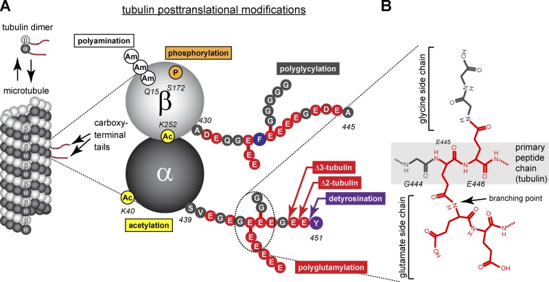
Microtubules are cytoskeletal filaments that are dynamically assembled from α/β-tubulin heterodimers. The primary sequence and structure of the tubulin proteins and, consequently, the properties and architecture of microtubules are highly conserved in eukaryotes. Despite this conservation, tubulin is subject to heterogeneity that is generated in two ways: by the expression of different tubulin isotypes and by posttranslational modifications (PTMs). Identifying the mechanisms that generate and control tubulin heterogeneity and how this heterogeneity affects microtubule function are long-standing goals in the field. Recent work on tubulin PTMs has shed light on how these modifications could contribute to a "tubulin code" that coordinates the complex functions of microtubules in cells.


Figures

Figure 1.
Figure 1.
Tubulin heterogeneity generated by PTMs. (A) Schematic representation of the distribution of different PTMs of tubulin on the α/β-tubulin dimer with respect to their position in the microtubule lattice. Acetylation (Ac), phosphorylation (P), and polyamination (Am) are found within the tubulin bodies that assemble into the microtubule lattice, whereas polyglutamylation, polyglycylation, detyrosination, and C-terminal deglutamylation take place within the C-terminal tubulin tails that project away from the lattice surface. The tubulin dimer represents TubA1A and TubB2B (Fig. 2), and modification sites for polyglutamylation and polyglycylation have been randomly chosen. (B) Chemical structure of the branched peptide formed by polyglutamylation and polyglycylation, using the γ-carboxyl groups of the modified glutamate residues as acceptor sites for the isopeptide bonds. Note that in the case of polyglutamylation, the elongation of the side chains generates classical peptide bonds (Redeker et al., 1991).
Figure 2.
Figure 2.
Heterogeneity of C-terminal tails of tubulin isotypes and their PTMs. The amino acid sequences of all tubulin genes found in the human genome are indicated, starting at the last amino acid of the folded tubulin bodies. Amino acids are represented in single-letter codes and color coded according to their biochemical properties. Known sites for polyglutamylation are indicated (Eddé et al., 1990; Alexander et al., 1991; Rüdiger et al., 1992). Potential modification sites (all glutamate residues) are indicated. Known C-terminal truncation reactions of α/β-tubulin (tub) are indicated. The C-terminal tails of the yeast Saccharomyces cerevisiae are shown to illustrate the phylogenetic diversity of these domains.
Figure 3.
Figure 3.
Enzymes involved in PTM of tubulin. Schematic representation of known enzymes (mammalian enzymes are shown) involved in the generation and removal of PTMs shown in Fig. 1. Note that some enzymes still remain unknown, and some modifications are irreversible. (*CCP5 preferentially removes branching points [Rogowski et al., 2010]; however, the enzyme can also hydrolyze linear glutamate chains [Berezniuk et al., 2013]).
Figure 4.
Figure 4.
Molecular components of the tubulin code. Schematic representation of potential coding elements that could generate specific signals for the tubulin code. (A) The length of the C-terminal tails of different tubulin isotypes differ significantly (Fig. 2) and could have an impact on the interactions between microtubules and MAPs. (B) Tubulin C-terminal tails are rich in charged amino acid residues. The distribution of these residues and local densities of charges could influence the electrostatic interactions with the tails and the readers. (C) Although each glutamate residue within the C-terminal tails could be considered a potential modification site, only some sites have been found highly occupied in tubulin purifications from native sources. This indicates selectivity of the modification reactions, which can participate in the generation of specific modification patterns (see D). Modification sites might be distinguished by their neighboring amino acid residues, which could create specific modification epitopes. (D) As a result of the large number of modification sites and the variability of side chains, a large variety of modification patterns could be generated within a single C-terminal tail of tubulin. (E) Modification patterns as shown in D can be distinct between α- and β-tubulin. These modification patterns could be differentially distributed at the surface of the microtubule lattice, thus generating a higher-order patterning. Tub, tubulin. For color coding, see Fig. 2.

References

    1. Aguilar, A., Becker L., Tedeschi T., Heller S., Iomini C., and Nachury M.V.. 2014. α-tubulin K40 acetylation is required for contact inhibition of proliferation and cell-substrate adhesion. Mol. Biol. Cell. 25:1854–1866 10.1091/mbc.E13-10-0609 - DOI - PMC - PubMed
    1. Aiken, J., Sept D., Costanzo M., Boone C., Cooper J.A., and Moore J.K.. 2014. Genome-wide analysis reveals novel and discrete functions for tubulin carboxy-terminal tails. Curr. Biol. 24:1295–1303 10.1016/j.cub.2014.03.078 - DOI - PMC - PubMed
    1. Akella, J.S., Wloga D., Kim J., Starostina N.G., Lyons-Abbott S., Morrissette N.S., Dougan S.T., Kipreos E.T., and Gaertig J.. 2010. MEC-17 is an alpha-tubulin acetyltransferase. Nature. 467:218–222 10.1038/nature09324 - DOI - PMC - PubMed
    1. Alexander, J.E., Hunt D.F., Lee M.K., Shabanowitz J., Michel H., Berlin S.C., MacDonald T.L., Sundberg R.J., Rebhun L.I., and Frankfurter A.. 1991. Characterization of posttranslational modifications in neuron-specific class III beta-tubulin by mass spectrometry. Proc. Natl. Acad. Sci. USA. 88:4685–4689 10.1073/pnas.88.11.4685 - DOI - PMC - PubMed
    1. Arce, C.A., and Barra H.S.. 1983. Association of tubulinyl-tyrosine carboxypeptidase with microtubules. FEBS Lett. 157:75–78 10.1016/0014-5793(83)81119-3 - DOI - PubMed

Publication types