. 2014 Aug 18;206(4):525-39.
doi: 10.1083/jcb.201404083.

Oxidoreductase activity is necessary for N-glycosylation of cysteine-proximal acceptor sites in glycoproteins

Affiliations

Oxidoreductase activity is necessary for N-glycosylation of cysteine-proximal acceptor sites in glycoproteins

Natalia A Cherepanova et al. J Cell Biol. .

Abstract

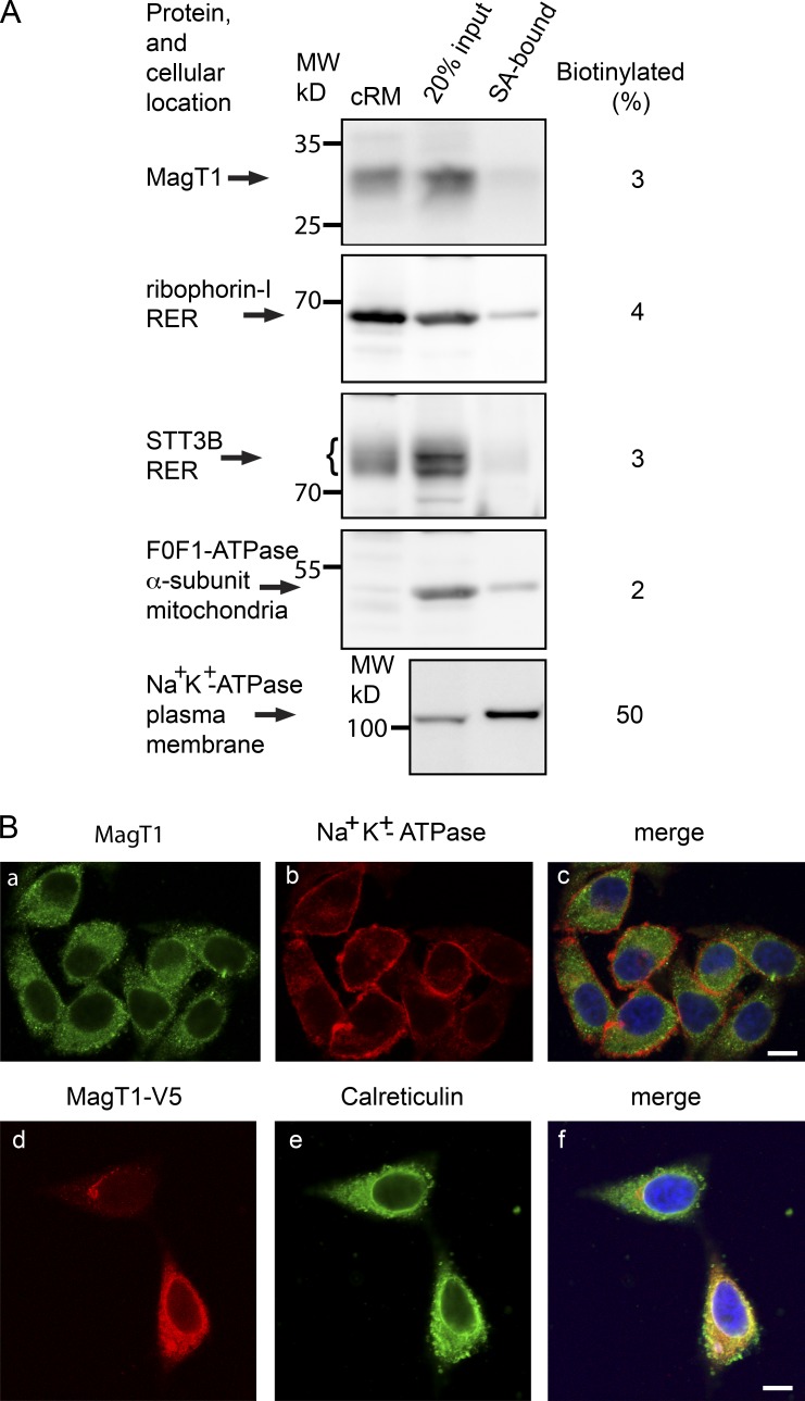
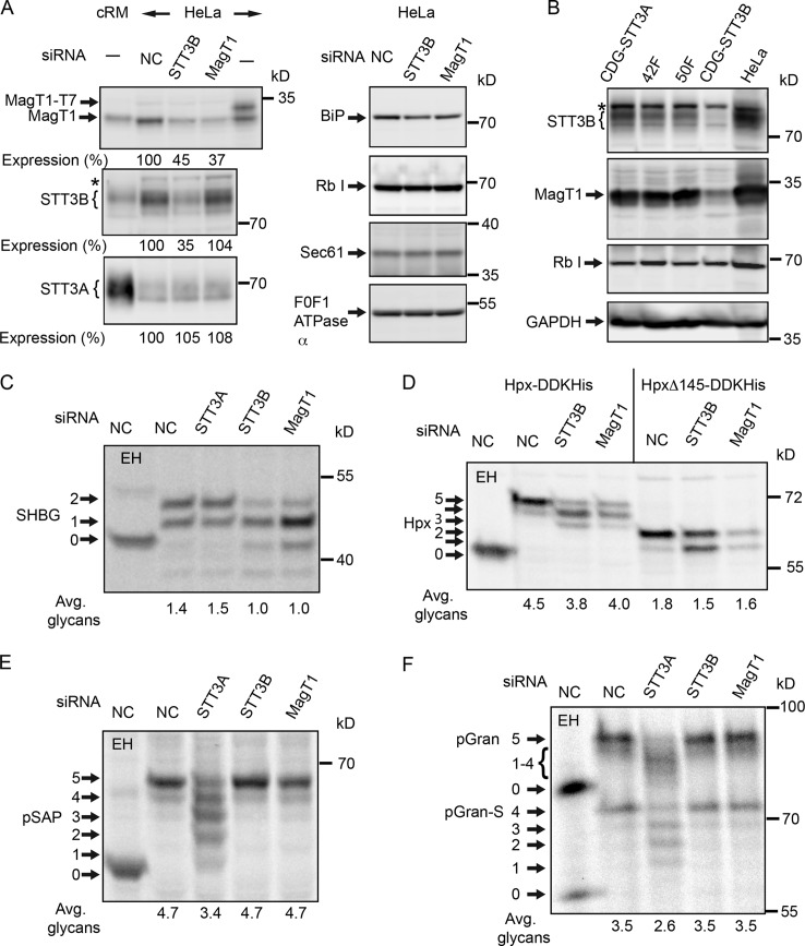
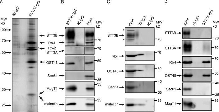
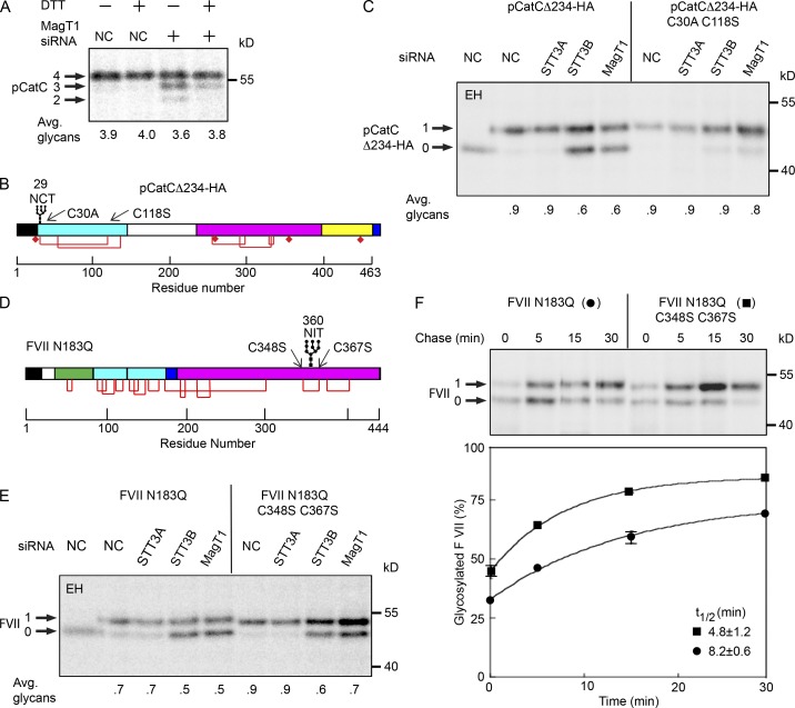
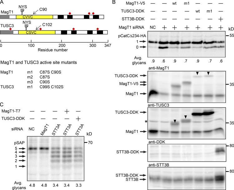
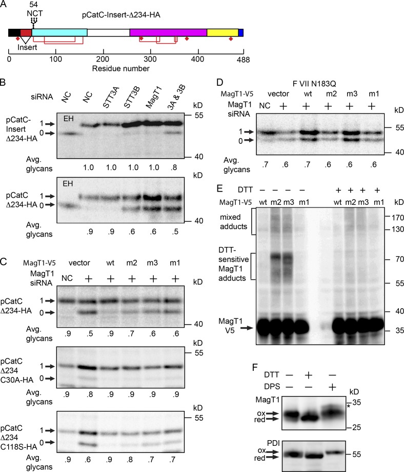
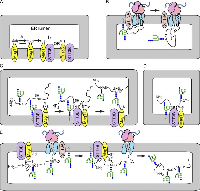
Stabilization of protein tertiary structure by disulfides can interfere with glycosylation of acceptor sites (NXT/S) in nascent polypeptides. Here, we show that MagT1, an ER-localized thioredoxin homologue, is a subunit of the STT3B isoform of the oligosaccharyltransferase (OST). The lumenally oriented active site CVVC motif in MagT1 is required for glycosylation of STT3B-dependent acceptor sites including those that are closely bracketed by disulfides or contain cysteine as the internal residue (NCT/S). The MagT1- and STT3B-dependent glycosylation of cysteine-proximal acceptor sites can be reduced by eliminating cysteine residues. The predominant form of MagT1 in vivo is oxidized, which is consistent with transient formation of mixed disulfides between MagT1 and a glycoprotein substrate to facilitate access of STT3B to unmodified acceptor sites. Cotranslational N-glycosylation by the STT3A isoform of the OST, which lacks MagT1, allows efficient modification of acceptor sites in cysteine-rich protein domains before disulfide bond formation. Thus, mammalian cells use two mechanisms to achieve N-glycosylation of cysteine proximal acceptor sites.


Figures

Figure 1.
Figure 1.
MagT1 is localized to the RER. (A) Cell-surface proteins labeled with sulfo-NHS-SS-biotin were isolated with streptavidin beads (SA-bound) and were resolved adjacent to sample input lanes by SDS-PAGE. The percentage of biotinylated protein was calculated as described in the Materials and methods. Canine pancreas RER membranes (cRM) were included as protein mobility standards for ER localized proteins. The indicated molecular weights shown in all figures are of prestained molecular weight markers electrophoresed on all gels. (B, a–c) HeLa cells were cultured for 24 h, fixed, and stained with antibodies to MagT1 (a and c, green) and the Na+K+-ATPase (b and c, red). (B, d–f) 24 h after transfection of HeLa cells with the MagT1-V5His expression vector, cells were fixed and stained with antibody against the V5 epitope (d and f, red) and calreticulin (e and f, green). The merged images (c and f) include the DAPI stain of the nucleus. Bars, 10 µm.
Figure 2.
Figure 2.
Hypoglycosylation of STT3B-dependent substrates in MagT1-depleted cells. (A and C–F) HeLa cells were treated with NC or siRNAs specific for STT3A, STT3B, or MagT1 for 72 h. (A) HeLa cell extracts and cRM were resolved by PAGE in SDS and analyzed by protein immunoblotting using the specified antisera. MagT1-T7 expressed in HeLa cells verified recognition of MagT1 by the anti-MagT1 sera. Expression values relative to cells treated with the NC siRNA are for the displayed image, which is representative of two or more experiments. (B) Cell extracts prepared from STT3A-CDG, STT3B-CDG, and normal control (42F and 50F) fibroblasts were resolved by PAGE in SDS and analyzed by protein immunoblotting. The F0F1-ATPase α (A) and glyceraldehyde 3-phosphate dehydrogenase (GAPDH; B) served as gel loading controls. The asterisks in A and B designate a nonspecific product recognized by the anti-STT3B sera. Protein expression levels for OST subunits were normalized to the F0F1-ATPase α subunit loading control and are expressed relative to the NC siRNA lane. (C–F) After 48 h of siRNA treatment, cells were transfected with expression vectors for SHBG (C), or Hpx (Hpx-DDKHis or HpxΔ145-DDKHis; D) and pulse-chase labeled (4 min pulse, 20 min chase) after an additional 24 h. (E and F) After 72 h of siRNA treatment, cells were pulse labeled for 4 min and chased for 10 min. As indicated, samples were digested with EH after immunoprecipitation with anti-SHBG (C), anti-DDK (D), anti-SapD (E), or anti-granulin (F). Glycoforms resolved by PAGE in SDS are labeled to indicate the number of N-linked glycans. EH-digested proteins migrate slightly slower than the nonglycosylated protein because of the presence of a single residual GlcNAc residue at each site. Quantified values below gel lanes (C–F) are for the displayed image, which is representative of two or more experiments.
Figure 3.
Figure 3.
MagT1 is a subunit of the STT3B complex. (A, B, and D) cRM were solubilized under nondenaturing conditions and incubated with protein A–Sepharose beads with covalently coupled nonimmune (NI) IgG or anti-STT3B IgG (A and B) or noncoupled anti-STT3A IgG (D, anti-ITM1 sera). (C) HeLa cells expressing MagT1-V5 were solubilized and incubated with protein A–Sepharose beads coated with anti-V5 IgG or NI IgG. (A–D) Proteins were eluted with IP wash buffer, resolved by SDS-PAGE, and stained with silver (A) to detect major proteins including known OST subunits (STT3B, ribophorin I [Rb-1], ribophorin II [Rb-II], and OST48) or analyzed by protein immunoblotting using the indicated antisera (B–D). (B–D) Input samples (cRM [B and D] or HeLa cell extract [C]) were electrophoresed on the same gel to provide protein mobility markers. MagT1 and malectin co-migrate (B) with the 34-kD band detected by silver staining (A). Asterisks designate nonspecific bands recognized by the anti-STT3B sera on protein blots of the input samples. In B, vertical lines indicate removal of an intervening lane of molecular weight markers.
Figure 4.
Figure 4.
Disulfide bonds in MagT1-dependent substrates. (A) HeLa cells treated with NC or MagT1 siRNA were treated with 3 mM DTT for 5 min before a 5-min pulse, 10-min chase labeling period. Endogenous cathepsin C was immunoprecipitated using anti-CatC sera and resolved by SDS-PAGE. Diagrams of pCatCΔ234-HA (B) and FVII N183Q (D) showing the signal sequence (black), glycosylation sites, disulfide bonds (red lines), free cysteine residues (diamonds), mature protein domains (green, cyan, magenta, and yellow segments), and the C-terminal HA tag on pCatCΔ234-HA. Disulfides that link (pCatCΔ234) or bracket (FVII N183Q) a STT3B-dependent glycosylation site are indicated. (C and E) HeLa cells were treated with NC, or siRNAs specific for STT3A, STT3B, or MagT1 for 48 h as indicated, then transfected with pCatCΔ234-HA (C) or FVII N183Q (E and F) expression vectors and cultured for an additional 24 h before pulse labeling. Cells were pulse labeled for 4 min (C), pulse labeled for 2 min, and chased for 30 min (E), or pulsed for 2 min and chased as indicated (F). Glycoprotein substrates were precipitated with anti-HA sera (C) or anti-factor VII sera (E and F). Quantified values below gel lanes (A, C, and E) are for the displayed image that is representative of two or more experiments. Data points in F are the mean of two determinations, with individual data points indicated by error bars.
Figure 5.
Figure 5.
Requirement for active site cysteine residues in MagT1 and TUSC3. (A) Diagrams of MagT1 and TUSC3 showing the N-terminal signal sequence (gray), lumenal thioredoxin domain (yellow) with active-site CXXC motif, glycosylation site, noncatalytic cysteine residues (red squares), and membrane spanning segments (black). Nomenclature for single and double cysteine mutants of MagT1 and TUSC3 is given. (B) HeLa cells were treated with NC or MagT1 siRNA for 48 h before cotransfection with a pCatCΔ234-HA expression vector and a wild-type or mutant MagT1-V5, TUSC3-DDK, or STT3B-DDK expression vector. Cells were pulse labeled for 4 min and chased for 10 min. Glycoforms of CatCΔ234-HA were collected by immunoprecipitation with anti-HA and resolved by PAGE in SDS. Total protein extracts from cells were resolved by SDS-PAGE and analyzed by protein immunoblotting using anti-MagT1, anti-TUSC3, anti-DDK, and anti-STT3B sera. Downward pointing arrowheads in the anti-MagT1 and anti-TUSC3 blots designate cross-reaction with TUSC3-DDK and MagT1-V5, respectively. The band designated by the asterisk is not TUSC3, but is a nonspecific background protein (see Fig. S2 B). (C) HeLa cells treated for 48 h with NC, STT3A siRNA, or MagT1 siRNA were transfected with MagT1-T7 or TUSC3-DDK expression vectors as indicated 24 h before pulse labeling for 4 min and chase for 10 min. Prosaposin glycoforms were immunoprecipitated using anti–saposin D sera and resolved by SDS-PAGE. Quantified values below gel lanes (B and C) are for the displayed image that is representative of two experiments.
Figure 6.
Figure 6.
Formation of mixed disulfides between MagT1 and glycoprotein substrates. (A) Diagram of the pCatC-Insert-Δ234 construct. (B–D) HeLa cells were treated with the NC or MagT1 siRNA for 48 h before cotransfection with wild-type or mutant versions of the pCatCΔ234-HA (B and C), FVII N183Q (D), and MagT1-V5 expression vectors (B–D). The m1, m2, and m3 mutants of MagT1 are defined in Fig. 5 A. Cells were pulse labeled for 4 min (A–D) and chased for 10 min (B and C) or 40 min (D). Glycoproteins were immunoprecipitated with anti-HA sera (B and C) or anti-FVII sera (D) and quantified after SDS-PAGE. (E) Cells expressing wild-type or mutant versions of MagT1-V5 were treated with NEM to prevent disulfide exchange during cell lysis and sample preparation. Total cell extracts were resolved by nonreducing (−DTT) or reducing (+DTT) SDS-PAGE as indicated, and analyzed by protein immunoblotting using anti-V5 sera. (F) In vivo redox status of MagT1 and PDI in HeLa cells was assayed using a maleimide-shift protocol. The arrows designate oxidized and reduced forms of MagT1 and PDI. A minor MagT1 reactive band in the DPS-oxidized lane (asterisk) is probably due to inefficient formation of a disulfide between cysteine residues located on the cytoplasmic face of TM3 and TM4 (see Fig. 5 A for a map of MagT1 cysteine residues). Quantified values below gel lanes (B–D) are for the displayed image, which is representative of two experiments.
Figure 7.
Figure 7.
MagT1-dependent glycosylation of sequons by the STT3B complex. (A) MagT1 or TUSC3, primarily in the oxidized state, assemble into the STT3B complex. (B) Cotranslational glycosylation of sequons in cysteine-rich protein domains by the STT3A complex. (C) Formation of a transient mixed disulfide between MagT1 and a glycoprotein substrate facilitates posttranslocational glycosylation of a cysteine-proximal sequon by the STT3B complex. (D) MagT1 is required for full activity of the STT3B complex even when substrates lack nearby cysteine residues. (E) The reduced form of MagT1, perhaps generated in situ, can reduce a disulfide by forming a transient mixed disulfide.

References

    1. Appenzeller-Herzog, C., and Ellgaard L.. 2008. In vivo reduction-oxidation state of protein disulfide isomerase: the two active sites independently occur in the reduced and oxidized forms. Antioxid. Redox Signal. 10:55–64 10.1089/ars.2007.1837 - DOI - PubMed
    1. Bas, T., Gao G.Y., Lvov A., Chandrasekhar K.D., Gilmore R., and Kobertz W.R.. 2011. Post-translational N-glycosylation of type I transmembrane KCNE1 peptides: implications for membrane protein biogenesis and disease. J. Biol. Chem. 286:28150–28159 10.1074/jbc.M111.235168 - DOI - PMC - PubMed
    1. Bocchinfuso, W.P., Ma K.L., Lee W.M., Warmels-Rodenhiser S., and Hammond G.L.. 1992. Selective removal of glycosylation sites from sex hormone-binding globulin by site-directed mutagenesis. Endocrinology. 131:2331–2336 - PubMed
    1. Bolt, G., Kristensen C., and Steenstrup T.D.. 2005. Posttranslational N-glycosylation takes place during the normal processing of human coagulation factor VII. Glycobiology. 15:541–547 10.1093/glycob/cwi032 - DOI - PubMed
    1. Bulleid, N.J., and Ellgaard L.. 2011. Multiple ways to make disulfides. Trends Biochem. Sci. 36:485–492 10.1016/j.tibs.2011.05.004 - DOI - PubMed

Publication types

MeSH terms