Review
. 2014 Sep 26;369(1652):20130505.
doi: 10.1098/rstb.2013.0505.

Extracellular-vesicle type of volume transmission and tunnelling-nanotube type of wiring transmission add a new dimension to brain neuro-glial networks

Affiliations
Review

Extracellular-vesicle type of volume transmission and tunnelling-nanotube type of wiring transmission add a new dimension to brain neuro-glial networks

Luigi F Agnati et al. Philos Trans R Soc Lond B Biol Sci. .

Abstract

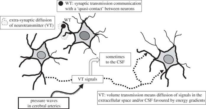
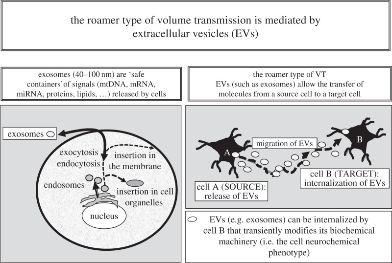
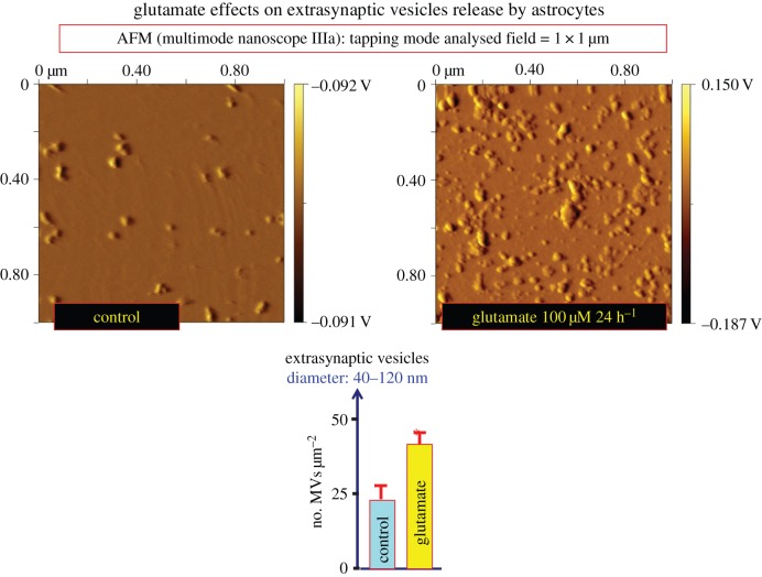
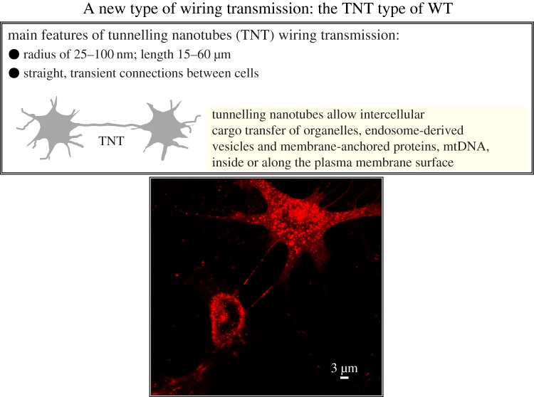
Two major types of intercellular communication are found in the central nervous system (CNS), namely wiring transmission (WT; point-to-point communication via private channels, e.g. synaptic transmission) and volume transmission (VT; communication in the extracellular fluid and in the cerebrospinal fluid). Volume and synaptic transmission become integrated because their chemical signals activate different types of interacting receptors in heteroreceptor complexes located synaptically and extrasynaptically in the plasma membrane. In VT, we focus on the role of the extracellular-vesicle type of VT, and in WT, on the potential role of the tunnelling-nanotube (TNT) type of WT. The so-called exosomes appear to be the major vesicular carrier for intercellular communication but the larger microvesicles also participate. Extracellular vesicles are released from cultured cortical neurons and different types of glial cells and modulate the signalling of the neuronal-glial networks of the CNS. This type of VT has pathological relevance, and epigenetic mechanisms may participate in the modulation of extracellular-vesicle-mediated VT. Gerdes and co-workers proposed the existence of a novel type of WT based on TNTs, which are straight transcellular channels leading to the formation in vitro of syncytial cellular networks found also in neuronal and glial cultures.

Keywords: exosomes; extracellular vesicles; microvesicles; tunnelling nanotubes; volume transmission; wiring transmission.


Figures

Figure 1.
Figure 1.
Schematic of wiring transmission and volume transmission in neural networks. Wiring transmission is illustrated by means of a synaptic contact. Volume transmission is the diffusion and flow of signals in the extracellular space (ECS) of the brain and in the cerebrospinal fluid (CSF) along the energy gradients. Thus, VT is illustrated as: the local extrasynaptic diffusion of neurotransmitters in the ECS; the long-distance diffusion and flow of some classical neurotransmitters and neuropeptides in the ECS; the flow of VT chemical signals via the CSF. Finally, the effects of the pressure waves in cerebral arteries on the diffusion and flow of the VT signals in the ECS and CSF are indicated (see [6,7,12,13]).
Figure 2.
Figure 2.
Schematic of some main aspects of the roamer type of VT and of its possible implications for integrative actions in the CNS. It was shown inter alia that (i) exosomes can mediate oligodendrocyte–neuron communication [77]; (ii) exosomes can play a role in interconnections between brain and peripheral organs since, for example, cardiac myocytes release exosomes [84] and exosomes can cross the blood–brain barrier [85]; and (iii) exosomes can cause transient cell phenotype changes. Thus, it was shown that exosomes allow intercellular transfer of GPCRs [53].
Figure 3.
Figure 3.
Effects of glutamate (100 μM) for 24 h on the release of extracellular vesicles from glioblastoma cultures (U87MG). Conditioned medium (33 ml) was collected and processed for exosome isolation. Extracellular vesicles were purified by differential centrifugation at 4°C, starting with a centrifugation at 300g (10 min) and followed by centrifugations at 12 000g for (20 min), and 100 000g (120 min). The resulting extracellular vesicle pellets were washed with phosphate-buffered saline (PBS) and then collected again by ultracentrifugation at 100 000g (120 min) and resuspended in 500 µl PBS. Purified exosomes were further diluted up to 1 : 150 ratio with PBS and evaluated for number and size by atomic force microscopy (AFM) analysis. In detail, 10 μl of the obtained suspension was adsorbed to freshly cleaved mica sheets for 15 min at room temperature, rinsed with deionized water, and air dried. A nanoscope IIIa multimode AFM (Veeco) in tapping mode with silicon probes (K ≈ 50 N m−1) was used. Constant force was maintained for imaging all samples. Topographic (height) and amplitude images were recorded simultaneously at 512 × 512 pixels at a scan rate of 2.03 Hz. The height and amplitude (equivalent to a map of the slope of the sample) images are representative of the exosome morphological characteristics. Height image processing was performed using Gwyddion 2.5 software. Data are presented as number of extracellular vesicles µm−2 ± s.e. (n = 9–12). The colour scale on the right expresses the height of the surface features in intrinsic units (voltage), representing the voltage generated by the piezoelectric cantilever when the tip is moved at each given z-coordinate. This voltage is linearly related to the height in natural coordinates (nm). Thus, the colour bar has as a minimum value of elevation, the brown colour, and as a maximum value of elevation, the white colour (L. F. Agnati, D. Guidolin, G. Maura, C.Tortorella, M. Marcoli, G. Leo, C. Carone, S. Genedani, D.O. Borroto-Escuela and K. Fuxe 2013, unpublished data). (Online version in colour.)
Figure 4.
Figure 4.
Upper panel: schematic of some main aspects of the tunnelling nanotubes (TNTs) WT [–34,107]. Lower panel: morphological evidence that TNTs can connect rat primary neuronal cells. The experimental procedure was as follows: TNT fluorescence labelling was performed by a fluorescent wheat germ agglutinin conjugate (WGA; Alexa Fluor 594 conjugate, 1 mg ml−1; Molecular Probes, Invitrogen, Eugene, OR, USA) directly added to the culture medium (1 : 400) at room temperature for 10 min. Subsequently, cells were washed with serum-free medium, and fresh medium was added. Live cell images were taken with a Leica DMIRE2 confocal microscope equipped with a 63× oil objective. (Online version in colour.)

References

    1. Agnati LF, Fuxe K, Zoli M, Ozini I, Toffano G, Ferraguti F. 1986. A correlation analysis of the regional distribution of central enkephalin and beta-endorphin immunoreactive terminals and of opiate receptors in adult and old male rats. Evidence for the existence of two main types of communication in the central nervous system: the volume transmission and the wiring transmission. Acta Physiol. Scand. 128, 201–207. (10.1111/j.1748-1716.1986.tb07967.x) - DOI - PubMed
    1. Agnati LF, Guidolin D, Baluska F, Leo G, Barlow PW, Carone C, Genedani S. 2010. A new hypothesis of pathogenesis based on the divorce between mitochondria and their host cells: possible relevance for Alzheimer's disease. Curr. Alzheimer Res. 7, 307–322. (10.2174/156720510791162395) - DOI - PubMed
    1. Fuxe K, Dahlstrom AB, Jonsson G, Marcellino D, Guescini M, Dam M, Manger P, Agnati L. 2010. The discovery of central monoamine neurons gave volume transmission to the wired brain. Prog. Neurobiol. 90, 82–100. (10.1016/j.pneurobio.2009.10.012) - DOI - PubMed
    1. Fuxe K, et al. 2007. From the Golgi–Cajal mapping to the transmitter-based characterization of the neuronal networks leading to two modes of brain communication: wiring and volume transmission. Brain Res. Rev. 55, 17–54. (10.1016/j.brainresrev.2007.02.009) - DOI - PubMed
    1. Fuxe K, Agnati LF, Zoli M, Cintra A, Harfstrand A, von Euler G, Grimaldi R, Kalia M, Eneroth P. 1988. The opioid peptide systems: their organization and role in volume transmission and neuroendocrine regulation. In Regulatory roles of opioid peptides (eds Illes P, Farsang C.), pp. 33–68. Weinheim, Germany: VCH.

Publication types