. 2014 Sep 26;369(1652):20130510.
doi: 10.1098/rstb.2013.0510.

Multifaceted effects of oligodendroglial exosomes on neurons: impact on neuronal firing rate, signal transduction and gene regulation

Affiliations

Multifaceted effects of oligodendroglial exosomes on neurons: impact on neuronal firing rate, signal transduction and gene regulation

Dominik Fröhlich et al. Philos Trans R Soc Lond B Biol Sci. .

Abstract

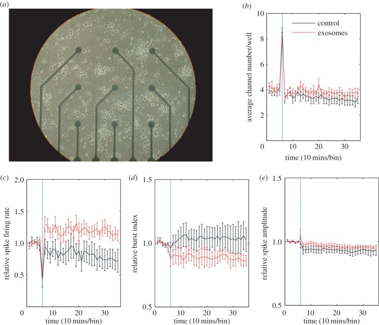
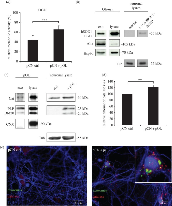
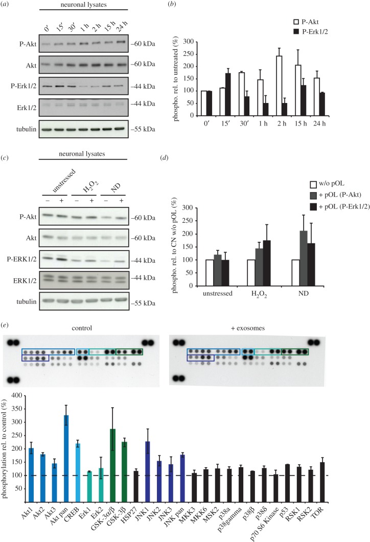
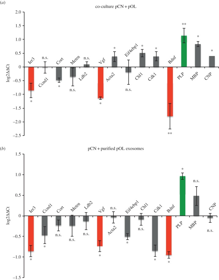
Exosomes are small membranous vesicles of endocytic origin that are released by almost every cell type. They exert versatile functions in intercellular communication important for many physiological and pathological processes. Recently, exosomes attracted interest with regard to their role in cell-cell communication in the nervous system. We have shown that exosomes released from oligodendrocytes upon stimulation with the neurotransmitter glutamate are internalized by neurons and enhance the neuronal stress tolerance. Here, we demonstrate that oligodendroglial exosomes also promote neuronal survival during oxygen-glucose deprivation, a model of cerebral ischaemia. We show the transfer from oligodendrocytes to neurons of superoxide dismutase and catalase, enzymes which are known to help cells to resist oxidative stress. Additionally, we identify various effects of oligodendroglial exosomes on neuronal physiology. Electrophysiological analysis using in vitro multi-electrode arrays revealed an increased firing rate of neurons exposed to oligodendroglial exosomes. Moreover, gene expression analysis and phosphorylation arrays uncovered differentially expressed genes and altered signal transduction pathways in neurons after exosome treatment. Our study thus provides new insight into the broad spectrum of action of oligodendroglial exosomes and their effects on neuronal physiology. The exchange of extracellular vesicles between neural cells may exhibit remarkable potential to impact brain performance.

Keywords: exosomes; extracellular vesicles; gene regulation; neuron–glia interaction; oligodendrocytes; signal transduction.


Figures

Figure 1.
Figure 1.
Impact of oligodendroglial exosomes on neuronal electrical activity. (a) Image of a 14 DIV pCN culture grown in a 9-channel MEA. Electrode spacing is 200 μm. (be) Recordings were performed at DIV 14. The blue vertical line indicates the application of pOL exosomes (red graph, n = 9) or sham control (black graph, n = 6) after 1 h of recording. Average channel number/well (b), relative spike firing rate (c), relative burst index (d) and relative spike amplitude (e) are depicted over 6 h of recording. Error bars ± s.d. (*p < 0.05; Students t-test).
Figure 2.
Figure 2.
Neuroprotection mediated by exosomes. (a) pCN exposed to pOL exosomes in Boyden chamber co-cultures and subjected to OGD compared to control conditions (pCN ctrl, pOL-conditioned medium depleted of exosomes). After OGD followed by reoxygenation, neuronal metabolic activity was determined by MTT assay. The relative metabolic activity correlates OGD co-cultures to respective cultures grown under normoxic conditions. Error bars, s.e.m. (***p < 0.001; n = 5, Students t-test). (bd) Western blot analysis of pCN lysates after co-culture with Oli-neu cells (ON) or pOL. (b) Presence of hSOD1-EGFP in isolated exosomes (left) and transfer of hSOD1-EGFP from transfected ON cells to pCN (right). Control pCN were co-cultured with untransfected ON. Alix and Hsp70 identify exosomes. (c) Western blotting of isolated pOL-derived exosomes demonstrates presence of catalase (Cat, left). PLP/DM20 serves as an oligodendrocyte-specific exosome marker and calnexin (CNX) as a contamination marker. Catalase levels determined after co-culture of pCN with pOL (right). Detection of PLP/DM20 in pCN indicates exosome transfer. (d) Quantification of catalase normalized to tubulin (Tub) as derived from Western blot signals in (c) (right). Error bars, s.e.m. (**p < 0.01; Wilcoxon test, n = 10). (e) Images of pCN co-cultured with pOL (right panel), which were stained with PKH67 to label pOL-derived exosomes (green). Control pCN were cultured in absence of pOL (left panel). pCN were immuno-stained for catalase (red) and the neuronal marker Tuj1 (blue). Scale bar, 20 µm.
Figure 3.
Figure 3.
Analysis of exosome-dependent signalling pathways in neurons. (a,b) Purified oligodendroglial exosomes were applied to pCN for 0 min, 15 min, 30 min, 1 h, 2 h, 15 h and 24 h. P-Akt and P-Erk1/2 levels determined by Western blotting were normalized to total Akt or Erk and expressed in relation to untreated pCN at timepoint 0 min (n = 2). (c,d) Analysis of exosome signalling in recipient neurons under stress conditions. pCN co-cultured in Boyden chambers with (+) or without (−) pOL for 48 h were subjected to oxidative stress (25 µM H2O2 for 1 h before lysis) or ND (culture in absence of B27 supplement during co-culture) followed by Western blot analysis compared to unstressed cells. (d) Relative P-Akt and P-Erk1/2 levels in pCN are expressed in relation to control cells (−) of each condition to selectively visualize exosome-dependent kinase activation (n = 3). (e) Phospho-MAPK Array for phosphorylated proteins detected in neuronal lysates. pCN were either treated with PBS (control) or purified oligodendroglial exosomes and subjected to oxidative stress (n = 4). Abundance of phosphorylated proteins was quantified by densitometry and compared to PBS-treated pCN (dashed line). Proteins showing a higher phosphorylation level compared to control are highlighted by coloured boxes. Error bars, s.e.m.
Figure 4.
Figure 4.
Validation of exosome-dependent neuronal gene regulation. qRT-PCR analysis of candidate genes selected from microarray analysis. (a) pCN co-cultured in Boyden chambers with pOL compared to control pCN (n = 6; BDNF and PLP, n = 14). (b) pCN treated with isolated pOL-derived exosomes compared to untreated pCN (n = 7; BDNF, PLP, MBP and CNP, n = 5). Pgk1 was used as normalization standard. Log2 values of the ΔΔCt values are displayed. Genes validated in both paradigms as downregulated or upregulated are indicated by red bars (dark grey in print) and green bars (light grey in print), respectively. Error bars, s.e.m. (n.s., not significant; *p < 0.05; **p < 0.01; Wilcoxon test). (Online version in colour.)
Figure 5.
Figure 5.
Downregulation of DCX in neurons in response to exosomes derived from oligodendrocyte precursor cells. (ac) Western blot analysis of neuronal lysates. (a) pCN were treated with exosomes isolated from Oli-neu cells ectopically expressing Sirt2-EYFP and PLP-EGFP for 2 and 24 h, respectively (n = 6). (b,c) Boyden chamber co-culture of pCN either with Oli-neu cells ectopically expressing Sirt-EYFP and PLP-EGFP (b, n = 4) or with pOL and Oli-neu (c, n = 6) for 2 days. (d) Doublecortin (DCX) expression levels normalized to tubulin (Tub) as quantified from Western blots. Control pCN (untreated) are referred to as 100%. Error bars, s.e.m. (*p < 0.05; **p < 0.01; ***p < 0.001; Students t-test).

References

    1. Raposo G, Stoorvogel W. 2013. Extracellular vesicles: exosomes, microvesicles, and friends. J. Cell Biol. 200, 373–383. (10.1083/jcb.201211138) - DOI - PMC - PubMed
    1. Johnstone RM, Mathew A, Mason AB, Teng K. 1991. Exosome formation during maturation of mammalian and avian reticulocytes: evidence that exosome release is a major route for externalization of obsolete membrane proteins. J. Cell Physiol. 147, 27–36. (10.1002/jcp.1041470105) - DOI - PubMed
    1. Ludwig AK, Giebel B. 2012. Exosomes: small vesicles participating in intercellular communication. Int. J. Biochem. Cell Biol. 44, 11–15. (10.1016/j.biocel.2011.10.005) - DOI - PubMed
    1. Bellingham SA, Guo BB, Coleman BM, Hill AF. 2012. Exosomes: vehicles for the transfer of toxic proteins associated with neurodegenerative diseases? Front. Physiol. 3, 124 (10.3389/fphys.2012.00124) - DOI - PMC - PubMed
    1. Thery C, Ostrowski M, Segura E. 2009. Membrane vesicles as conveyors of immune responses. Nat. Rev. Immunol. 9, 581–593. (10.1038/nri2567) - DOI - PubMed

Publication types

MeSH terms